Review
. 2020 Jun 26;21(12):4546.
doi: 10.3390/ijms21124546.

Betacoronavirus Genomes: How Genomic Information has been Used to Deal with Past Outbreaks and the COVID-19 Pandemic

Affiliations
Review

Betacoronavirus Genomes: How Genomic Information has been Used to Deal with Past Outbreaks and the COVID-19 Pandemic

Alejandro Llanes et al. Int J Mol Sci. .

Abstract

In the 21st century, three highly pathogenic betacoronaviruses have emerged, with an alarming rate of human morbidity and case fatality. Genomic information has been widely used to understand the pathogenesis, animal origin and mode of transmission of coronaviruses in the aftermath of the 2002-2003 severe acute respiratory syndrome (SARS) and 2012 Middle East respiratory syndrome (MERS) outbreaks. Furthermore, genome sequencing and bioinformatic analysis have had an unprecedented relevance in the battle against the 2019-2020 coronavirus disease 2019 (COVID-19) pandemic, the newest and most devastating outbreak caused by a coronavirus in the history of mankind. Here, we review how genomic information has been used to tackle outbreaks caused by emerging, highly pathogenic, betacoronavirus strains, emphasizing on SARS-CoV, MERS-CoV and SARS-CoV-2. We focus on shared genomic features of the betacoronaviruses and the application of genomic information to phylogenetic analysis, molecular epidemiology and the design of diagnostic systems, potential drugs and vaccine candidates.

Keywords: COVID-19; MERS-CoV; SARS-CoV; SARS-CoV-2; betacoronaviruses; genomics.


Conflict of interest statement

The authors declare no conflict of interest.

Figures

Figure 1
Figure 1
Organization of betacoronavirus genomes. Name abbreviations are provided in Table 1.
Figure 2
Figure 2
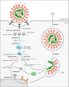
Replication cycle of a typical coronavirus. Upon recognition of the host cell receptor, the viral particle enters the host cell and is uncoated, releasing its positive-sense genomic RNA. Host ribosomes translate polyproteins pp1a and pp1ab, which self-cleave to produce the nonstructural proteins (Nsps). Several Nsps assemble into the replicase-transcriptase complex (RTC) that generates the mRNAs for structural and accessory proteins through transcription, as well as positive-sense genomic RNAs through replication. Viral core particles are assembled within smooth vesicles derived from the endoplasmic reticulum-Golgi intermediate compartment (ERGIC). The viral progeny is ultimately released via exocytosis.
Figure 3
Figure 3
Main functional domains in protein-coding genes. (A) Location of Nsps along the sequence of ORF1a and ORF1b. (B) Functional domains of Nsp3, Nsp5, Nsp12, Nsp13, Nsp14, Nsp15 and Nsp16. (C) Functional domains of structural proteins. All proteins are from SARS-CoV, except for the Nsp3 and HE proteins of murine hepatitis virus (MHV), which are included for comparative purposes. Proteins are drawn to scale, except for E and M, which are drawn three (3×) and two (2×) times larger, respectively. Specific domain name abbreviations are explained in the main text. TM: transmembrane domain, SP: signal peptide, FP: fusion peptide, RBD: receptor-binding domain, CP: cytoplasmic domain, NTD: N-terminal domain, CTD: C-terminal domain.
Figure 4
Figure 4
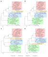
Phylogenetic analysis of representative betacoronaviruses. Figure shows four alternative phylogenies for coronaviruses in Table 1, inferred from complete genome sequences (A), concatenated sequences of ORF1ab domains (B), whole S protein (C) and the receptor-binding domain (RBD) (D). Phylogenetic analysis was performed as previously described [4,52], briefly, sequences were aligned with MAFFT [56] and trees were built with IQ-TREE [57], with the maximum likelihood (ML) method and the GTR+G+I model. For protein sequences, amino acid alignments were converted to nucleotides with PAL2NAL [58]. Numbers above or below branches indicate branch support measures expressed as percentage and estimated using the Shimodaira-Hasegawa (SH)-like approximate likelihood ratio test (aLRT) with 1000 replicates. Trees were rooted with human alphacoronaviruses 229E and NL63 (GenBank accession numbers NC_002645 and NC_005831, respectively).

References

    1. Cui J., Li F., Shi Z.L. Origin and evolution of pathogenic coronaviruses. Nat. Rev. Microbiol. 2019;17:181–192. doi: 10.1038/s41579-018-0118-9. - DOI - PMC - PubMed
    1. Sola I., Almazán F., Zúñiga S., Enjuanes L. Continuous and Discontinuous RNA Synthesis in Coronaviruses. Annu. Rev. Virol. 2015;2:265–288. doi: 10.1146/annurev-virology-100114-055218. - DOI - PMC - PubMed
    1. Li W., Moore M.J., Vasllieva N., Sui J., Wong S.K., Berne M.A., Somasundaran M., Sullivan J.L., Luzuriaga K., Greeneugh T.C., et al. Angiotensin-converting enzyme 2 is a functional receptor for the SARS coronavirus. Nature. 2003;426:450–454. doi: 10.1038/nature02145. - DOI - PMC - PubMed
    1. Zhou P., Yang X.L., Wang X.G., Hu B., Zhang L., Zhang W., Si H.R., Zhu Y., Li B., Huang C.L., et al. A pneumonia outbreak associated with a new coronavirus of probable bat origin. Nature. 2020;579:270–273. doi: 10.1038/s41586-020-2012-7. - DOI - PMC - PubMed
    1. Raj V.S., Mou H., Smits S.L., Dekkers D.H.W., Müller M.A., Dijkman R., Muth D., Demmers J.A.A., Zaki A., Fouchier R.A.M., et al. Dipeptidyl peptidase 4 is a functional receptor for the emerging human coronavirus-EMC. Nature. 2013;495:251–254. doi: 10.1038/nature12005. - DOI - PMC - PubMed

MeSH terms

LinkOut - more resources