Review
. 2020 Jun 26;9(6):1553.
doi: 10.3390/cells9061553.

Amyloid Proteins and Peripheral Neuropathy

Affiliations
Review

Amyloid Proteins and Peripheral Neuropathy

Mohammed M H Asiri et al. Cells. .

Abstract

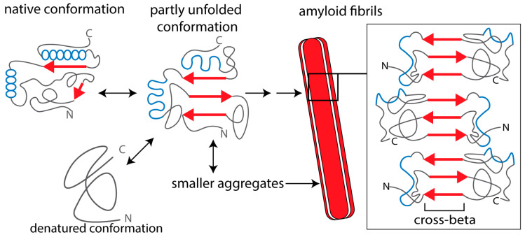
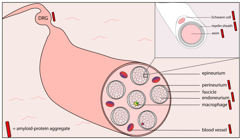
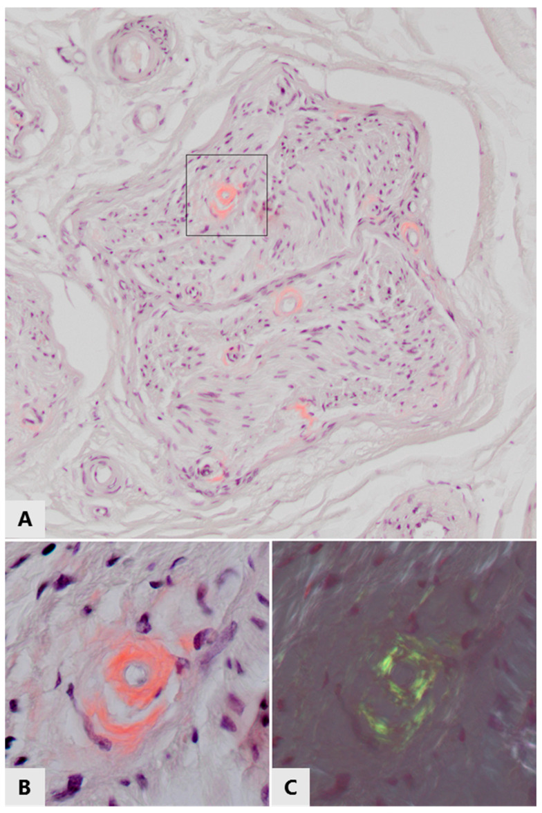
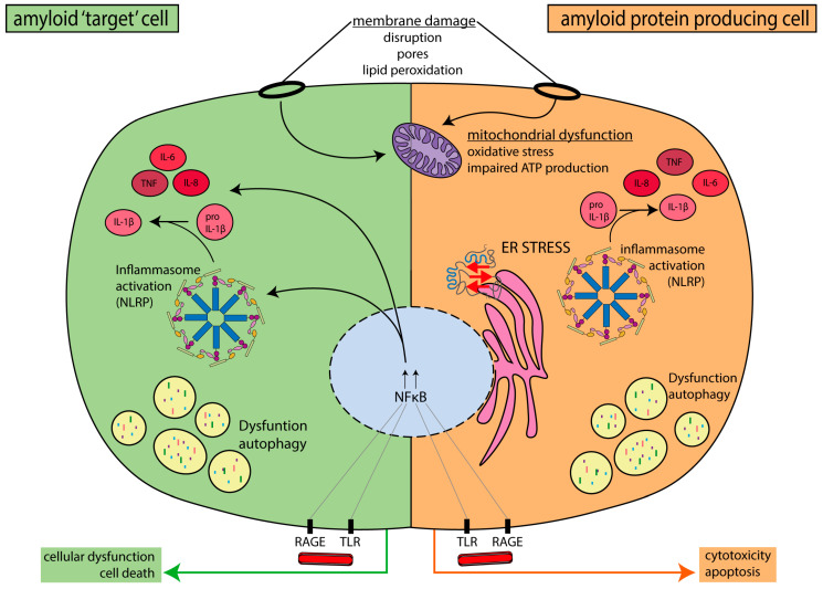
Painful peripheral neuropathy affects millions of people worldwide. Peripheral neuropathy develops in patients with various diseases, including rare familial or acquired amyloid polyneuropathies, as well as some common diseases, including type 2 diabetes mellitus and several chronic inflammatory diseases. Intriguingly, these diseases share a histopathological feature-deposits of amyloid-forming proteins in tissues. Amyloid-forming proteins may cause tissue dysregulation and damage, including damage to nerves, and may be a common cause of neuropathy in these, and potentially other, diseases. Here, we will discuss how amyloid proteins contribute to peripheral neuropathy by reviewing the current understanding of pathogenic mechanisms in known inherited and acquired (usually rare) amyloid neuropathies. In addition, we will discuss the potential role of amyloid proteins in peripheral neuropathy in some common diseases, which are not (yet) considered as amyloid neuropathies. We conclude that there are many similarities in the molecular and cell biological defects caused by aggregation of the various amyloid proteins in these different diseases and propose a common pathogenic pathway for "peripheral amyloid neuropathies".

Keywords: amyloid neuropathies; amyloid proteins; amyloidosis; chronic pain; peripheral neuropathy; type 2 diabetes mellitus.


Conflict of interest statement

The authors declare no conflicts of interest.

Figures

Figure 1
Figure 1
Schematic representation of amyloid protein aggregation. The β-strands in the amyloid-forming protein are indicated as red arrows and α-helices as blue spheres. In the native conformation, β-strands of the monomeric protein (if present) are not aligned and “shielded”, which prevents intermolecular aggregation. A partially unfolded or misfolded molecule can form different kinds of intermolecular aggregates. Amyloid oligomers are relatively small, compact structures that may be composed of antiparallel β-strands or contain α-helical conformations. Protofibrils and mature amyloid fibrils are formed via β-strand stacking, forming extended networks of β-sheets with a characteristic cross-beta structure. Mature fibrils consist of a few identical fibrillar “subunits”. Smaller aggregates (oligomers and protofibrils) are mostly cytotoxic, whereas extracellular, fibrillar amyloid deposits can also impair tissue and organ function by impairing blood supply to the cells (see text for references).
Figure 2
Figure 2
Schematic representation of the ultrastructure of a peripheral sensory nerve, with locations where amyloid or amyloid protein aggregates have been demonstrated indicated by a red bar (based on references [73,74,75,76,77,78,79,80,81]). DRG = dorsal root ganglion.
Figure 3
Figure 3
Amyloid deposits in/around small endoneurial blood vessels in the left sural nerve of a 70-year old patient with immunoglobulin light chain amyloidosis. (A) White light microscopy of Congo red stained section, showing pink-stained thickening of vascular walls in a nerve fasciculus. These thickened vascular walls also stained positive for lambda light chains (not shown). (B,C) Enlargement of the framed area of the top panel, (B) viewed with white light, (C) viewed with polarized light, showing green/yellow birefringence of the Congo red positive vascular walls, proving the amyloid nature of these light chain deposits.
Figure 4
Figure 4
Overview of the cell biological mechanisms that have been implicated in amyloid-protein-induced cellular damage and apoptosis and peripheral neuropathy, both in cells producing an amyloid protein and in other cells affected by amyloid or amyloid protein aggregates. Such “amyloid target cells” include cell types that degrade amyloid and protein aggregates after phagocytosis (macrophages and microglia), as well as Schwann cells (involved in nerve function and integrity) and endothelial cells (involved in microangiopathy).

References

    1. Collins M.P., Dyck P.J.B. Peripheral Nervous System Involvement. Rare Diseases of the Immune System. In: Sinico R., Guillevin L., editors. Anti-Neutrophil Cytoplasmic Antibody (ANCA) Associated Vasculitis. Springer; Cham, Switzerland: 2020.
    1. Scholz J., Finnerup N.B., Attal N., Aziz Q., Baron R., Bennett M.I., Benoliel R., Cohen M., Cruccu G., Davis K.D., et al. The IASP classification of chronic pain for ICD-11: Chronic neuropathic pain. Pain. 2019;160:53–59. doi: 10.1097/j.pain.0000000000001365. - DOI - PMC - PubMed
    1. Feldman E.L., Callaghan B.C., Pop-Busui R., Zochodne D.W., Wright D.E., Bennett D.L., Bril V., Russell J.W., Viswanathan V. Diabetic neuropathy. Nat. Rev. Dis. Primers. 2019;5:41. doi: 10.1038/s41572-019-0092-1. - DOI - PubMed
    1. Javed S., Hayat T., Menon L., Alam U., Malik R.A. Diabetic peripheral neuropathy in people with type 2 diabetes: Too little too late. Diabetic Med. 2019 doi: 10.1111/dme.14194. - DOI - PubMed
    1. Colloca L., Ludman T., Bouhassira D., Baron R., Dickenson A.H., Yarnitsky D., Freeman R., Truini A., Attal N., Finnerup N.B., et al. Neuropathic pain. Nat. Rev. Dis. Primers. 2017;3:17002. doi: 10.1038/nrdp.2017.2. - DOI - PMC - PubMed

Publication types

MeSH terms

Substances