Review
. 2020 Jun 26;10(6):1244.
doi: 10.3390/nano10061244.

Multi-Scale Surface Treatments of Titanium Implants for Rapid Osseointegration: A Review

Affiliations
Review

Multi-Scale Surface Treatments of Titanium Implants for Rapid Osseointegration: A Review

Qingge Wang et al. Nanomaterials (Basel). .

Abstract

The propose of this review was to summarize the advances in multi-scale surface technology of titanium implants to accelerate the osseointegration process. The several multi-scaled methods used for improving wettability, roughness, and bioactivity of implant surfaces are reviewed. In addition, macro-scale methods (e.g., 3D printing (3DP) and laser surface texturing (LST)), micro-scale (e.g., grit-blasting, acid-etching, and Sand-blasted, Large-grit, and Acid-etching (SLA)) and nano-scale methods (e.g., plasma-spraying and anodization) are also discussed, and these surfaces are known to have favorable properties in clinical applications. Functionalized coatings with organic and non-organic loadings suggest good prospects for the future of modern biotechnology. Nevertheless, because of high cost and low clinical validation, these partial coatings have not been commercially available so far. A large number of in vitro and in vivo investigations are necessary in order to obtain in-depth exploration about the efficiency of functional implant surfaces. The prospective titanium implants should possess the optimum chemistry, bionic characteristics, and standardized modern topographies to achieve rapid osseointegration.

Keywords: macro-scale; micro-scale; nano-scale; rapid bone integration; roughness; surface modification.


Conflict of interest statement

The authors declare no conflict of interest.

Figures

Figure 1
Figure 1
Structure of skeleton: descending hierarchical macro- to nano-scale structures of natural bone. (Reproduced with permission from [62]. Copyright Elsevier, 2016).
Figure 2
Figure 2
(a) Schematics of a laser beam melting (LBM) machine; (bd) porous structures; (ef) micro-CT images of human cancellous bones; (g) stacked hollow cubes; (hi) the surgical template in pre-blasting and after blasting condition; (j) the dental restorations; (k) a personalized femoral component. (Reproduced with permission from [93]. Copyright Elsevier, 2019).
Figure 3
Figure 3
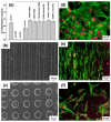
(a) XTT (Dimethoxazole yellow) results of cell viability of MC3T3-E1 fibroblast cells after 24 h in contact with the extracts in the as-received and laser textured surface; (b,c) SEM of the surface of linear geometry and dimple geometry; (df) fluorescent micrographs of the as-received, line geometry and dimple geometry showing the attachment of MC3T3-E1 cells. (Reproduced with permission from [126]. Copyright Elsevier, 2015).
Figure 4
Figure 4
(a) Standard version (grit-blasting); (b) HA coating on standard version; (c) straight Alloclassic™ THA in a 57-year-old female patient for nonunion of a fracture of the femoral neck after removing fixation hardware: immediate postoperative control; (d) radiographic result after 23 years and 3 months of follow-up at the age of 80 years old and 5 months. (Reproduced with permission from [145]. Copyright Elsevier, 2014).
Figure 5
Figure 5
Typical dental implant surface morphology using acid-etching. (Reproduced with permission from [161]. Copyright Elsevier, 2013).
Figure 6
Figure 6
(a) Representative histological images of 3D, 3DA, and SLA implants after implantation for 3 and 6 weeks, respectively (scale bar = 200 μm); (b) quantification of BIC percentages on implant surfaces; (c) SEM of 3D, 3DA, and SLA surfaces; (d) cell morphology on the 3DA surface after culturing of bone marrow stromal cells (BMSCs) for 24 h observed using SEM. (Reproduced with permission from [167]. Copyright Elsevier, 2020).
Figure 7
Figure 7
(a) Schematics during laser engineered net shaping (LENSTM) and plasma-spraying treatments; (b) bond strength between HA and HA/LENS coatings and substrates; (c) accumulative Ag+ release in MgO-Ag2O-HA/Ti-6Al-4V and MgO-Ag2O-HA/LENS/Ti-6Al-4V group. (Reproduced with permission from [194]. Copyright Elsevier, 2019).
Figure 8
Figure 8
(a) Schematics during anodization; (b) FESEM micrographs showing the morphology of the highly ordered TiO2 nanotubes present on NT-Ca/P; (c) high resolution XPS spectra of deconvoluted Ca 2p; (d) potentiodynamic polarization curves of Ti, NT, NT-Ca/P, and NT-RP-Ca/P samples immersed at 37 °C; (e) FESEM micrographs of MG-63 cells cultured on NT-RP-Ca/P surfaces after one day of incubation. (Reproduced with permission from [198]. Copyright Elsevier, 2016).

Similar articles

Cited by

References

    1. Guéhennec L.L., Soueidan A., Layrolle P., Inserm Y.A. Surface treatments of titanium dental implants for rapid osseointegration. Dent. Mater. 2006;3:844–854. doi: 10.1016/j.dental.2006.06.025. - DOI - PubMed
    1. Xiao L., Wu S., Liu B., Yang H., Yang L. Advances in metallic biomaterials with both osteogenic and anti-infection properties. ACTA Metall. Sin. 2017;53:1284–1302.
    1. Spriano S., Yamaguchi S., Baino F., Ferraris S. A critical review of multifunctional titanium surfaces: New frontiers for improving osseointegration and host response, avoiding bacteria contamination. Acta Biomater. 2018;79:1–22. doi: 10.1016/j.actbio.2018.08.013. - DOI - PubMed
    1. Brånemark P.-I., Breine U., Adell R., Hansson B.O., Lindström J., Ohlsson Å. Intra-osseous anchorage of dental prostheses: I. Experimental studies. Scand. J. Plast. Reconstr. Surg. 1969;3:81–100. doi: 10.3109/02844316909036699. - DOI - PubMed
    1. Chrcanovic B.R., Kisch J., Albrektsson T., Wennerberg A. A retrospective study on clinical and radiological outcomes of oral implants in patients followed up for a minimum of 20 years. Clin. Implant Dent. Relat. Res. 2018;20:199–207. doi: 10.1111/cid.12571. - DOI - PubMed

LinkOut - more resources