. 2020 Jun 26;8(6):176.
doi: 10.3390/biomedicines8060176.

Hyperthermia Disturbs and Delays Spontaneous Differentiation of Human Embryoid Bodies

Affiliations

Hyperthermia Disturbs and Delays Spontaneous Differentiation of Human Embryoid Bodies

Ji Hyun Kwon et al. Biomedicines. .

Abstract

Various types of stress stimuli have been shown to threaten the normal development of embryos during embryogenesis. Prolonged heat exposure is the most common stressor that poses a threat to embryo development. Despite the extensive investigation of heat stress control mechanisms in the cytosol, the endoplasmic reticulum (ER) heat stress response remains unclear. In this study, we used human embryonic stem cells (hESCs) to examine the effect of heat stress on early embryonic development, specifically alterations in the ER stress response. In a hyperthermic (42 °C) culture, ER stress response genes involved in hESC differentiation were induced within 1 h of exposure, which resulted in disturbed and delayed differentiation. In addition, hyperthermia increased the expression levels of activating transcription factor 4 (ATF4) and C/EBP homologous protein (CHOP) genes, which are associated with the protein kinase RNA-like endoplasmic reticulum kinase (PERK) signaling pathway. Furthermore, we demonstrated that tauroursodeoxycholic acid, a chemical chaperone, mitigated the delayed differentiation under hyperthermia. Our study identified novel gene markers in response to hyperthermia-induced ER stress on hESCs, thereby providing further insight into the mechanisms that regulate human embryogenesis.

Keywords: embryogenesis; endoplasmic reticulum; human embryonic stem cells; hyperthermia; stress response.


Conflict of interest statement

The authors declare no conflicts of interest.

Figures

Figure 1
Figure 1
Determination of the optimal experimental hyperthermic conditions for unfolded protein response (UPR) activation without causing human embryonic stem cell (hESC) death. (A) Phase-contrast images showing in vitro spontaneous differentiation of the hESCs under normal and hyperthermic conditions. (B) Gene expression analysis by RT-qPCR using total RNA isolated from spontaneously differentiated EBs exposed to a hyperthermic condition for the indicated times. The embryoid bodies (EBs) expressed UPR genes and stemness genes. Fold changes of signal intensity were normalized by GAPDH intensities. Fold changes represent the mean values obtained from the three independent experiments. Statistical significance was determined by comparing the relative band intensities referring to those of undifferentiated hESCs as 1. Student’s t-test: * p < 0.05, ** p < 0.01, and *** p < 0.001, **** p < 0.0001.
Figure 2
Figure 2
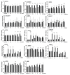
Hyperthermia activates the embryoid body (EB) stress pathway in human EBs. RT-qPCR analysis of the EBs cultured under normothermic (black bars) and hyperthermic (gray bars) conditions. Gene expression of the markers of stemness (OCT4, NANOG, and SOX2), lineage-specific markers (NeuroD1, BMP4, and SOX17), and ER stress-induced UPR-related genes (ATF4, ATF3, GADD34, CHOP, tXBP1, sXBP1, ATF6, and BIP). The housekeeping gene GAPDH was used as a loading control. Fold changes of signal intensity were normalized by GAPDH intensities. Fold changes shown are the mean values obtained from three independent experiments. Statistical significance was determined by comparing the relative band intensities and referring to those of undifferentiated hESCs as 1. Student’s t-test: * p < 0.05, ** p < 0.01, and *** p < 0.001, **** p < 0.0001.
Figure 3
Figure 3
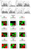
Hyperthermia disturbs spontaneously differentiating human EBs (hEBs) through UPR induction. (A) Phase-contrast images showing the in vitro spontaneous differentiation of the hESCs for the indicated days after exposure to hyperthermia or normothermia for 4 h. (B) Heatmap of the differentiation potential of all the gene classes (mesoderm, endoderm, ectoderm, mesendoderm, and self-renewal) of EBs at day 12 after exposure to hyperthermia or normothermia for 4 h. (C) Expression of pluripotency markers (OCT4, NANOG, and SOX2) and differentiation markers (ectoderm: NeuroD1, NR2F2, PAX6; mesoderm: Hand1, SMA, BMP4; endoderm: GATA6, AFP, SOX17) examined by RT-qPCR. The black bars indicate the normal condition and the gray bars indicate the hyperthermia condition. The housekeeping gene GAPDH was used as a loading control. Fold changes in the signal intensity were normalized with GAPDH. Fold changes shown are the mean values obtained from three independent experiments. Statistical significance was determined by comparing the relative band intensities and referring to those of undifferentiated hESCs as 1. Student’s t-test: * p < 0.05, ** p < 0.01, and *** p < 0.001, **** p < 0.0001.
Figure 4
Figure 4
Tauroursodeoxycholic acid (TUDCA) suppressed the induction of UPR-related genes under heat stress. (A) UPR-related genes measured by RT-qPCR. Left: EBs were cultured under normal conditions for the indicated times; right: DMSO (control)- or TUDCA-treated EBs cultured under hyperthermic conditions for the indicated times. Fold changes shown are the mean values obtained from three independent experiments. Statistical significance was determined by comparing the relative band intensities with those of undifferentiated hESCs as 1. Student’s t-test: * p < 0.05, ** p < 0.01, and *** p < 0.001, **** p < 0.0001. The Black bars indicate the normal condition, black border gray bars indicate the hyperthermic condition, and gray bars indicate the hyperthermic condition with TUDCA. (B) Heatmap of the differentiation markers (ectoderm: OLFM3, NR2F2, NeuroD1; mesoderm: SMA, TBX3, BMP4; endoderm: GATA6, AFP, SOX17) expressed over time from day 0 to day 12.

References

    1. Ziskin M.C., Morrissey J. Thermal Thresholds for Teratogenicity, Reproduction, and Development. Int. J. Hyperth. 2011;27:374–387. doi: 10.3109/02656736.2011.553769. - DOI - PubMed
    1. Zou J., Guo Y., Guettouche T., Smith D.F., Voellmy R. Repression of heat shock transcription factor HSF1 activation by HSP90 (HSP90 complex) that forms a stress-sensitive complex with HSF1. Cell. 1998;94:471–480. doi: 10.1016/S0092-8674(00)81588-3. - DOI - PubMed
    1. Anckar J., Sistonen L. Regulation of HSF1 function in the heat stress response: Implications in aging and disease. Annu. Rev. Biochem. 2011;80:1089–1115. doi: 10.1146/annurev-biochem-060809-095203. - DOI - PubMed
    1. Guo Y., Guettouche T., Fenna M., Boellmann F., Pratt W.B., Toft D.O., Smith D.F., Voellmy R. Evidence for a mechanism of repression of heat shock factor 1 transcriptional activity by a multichaperone complex. J. Biol. Chem. 2001;276:45791–45799. doi: 10.1074/jbc.M105931200. - DOI - PubMed
    1. Park S., Lim Y., Lee D., Elvira R., Lee J.M., Lee M.R., Han J. Modulation of Protein Synthesis by eIF2alpha Phosphorylation Protects Cell from Heat Stress-Mediated Apoptosis. Cells. 2018;7:254. doi: 10.3390/cells7120254. - DOI - PMC - PubMed