Review
. 2020 Jun 26;11(6):711.
doi: 10.3390/genes11060711.

Reciprocal Regulation between Primary Cilia and mTORC1

Affiliations
Review

Reciprocal Regulation between Primary Cilia and mTORC1

Yandong Lai et al. Genes (Basel). .

Abstract

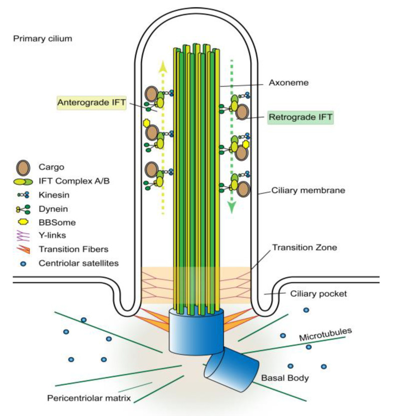
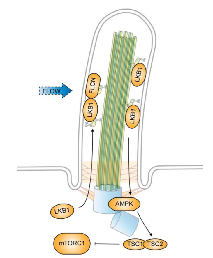
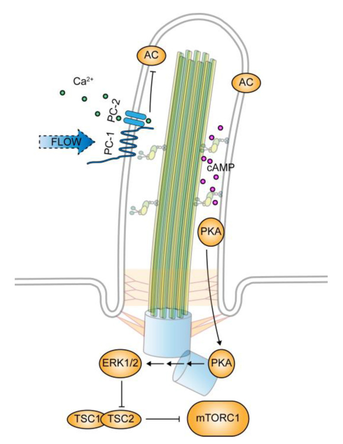
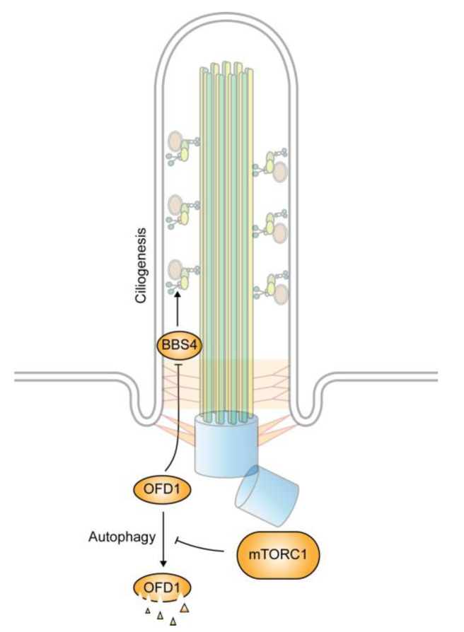
In quiescent cells, primary cilia function as a mechanosensor that converts mechanic signals into chemical activities. This unique organelle plays a critical role in restricting mechanistic target of rapamycin complex 1 (mTORC1) signaling, which is essential for quiescent cells to maintain their quiescence. Multiple mechanisms have been identified that mediate the inhibitory effect of primary cilia on mTORC1 signaling. These mechanisms depend on several tumor suppressor proteins localized within the ciliary compartment, including liver kinase B1 (LKB1), AMP-activated protein kinase (AMPK), polycystin-1, and polycystin-2. Conversely, changes in mTORC1 activity are able to affect ciliogenesis and stability indirectly through autophagy. In this review, we summarize recent advances in our understanding of the reciprocal regulation of mTORC1 and primary cilia.

Keywords: AMPK; LKB1; Tsc2; autophagy; ciliogenesis; mTOR; mTORC1; polycystin-1; primary cilia.


Conflict of interest statement

The authors declare no conflict of interest.

Figures

Figure 1
Figure 1
Structure of the primary cilium. The primary cilium is a solitary cell surface protrusion composed of a microtubule-based core (axoneme) ensheathed by an extension of the plasma membrane. The axoneme contains nine pairs microtubules emanating from the basal body located at the base of the cilium. The proximal ends of axonemal microtubules are tethered to the ciliary membrane by Y-links, which form the transition zone. The ciliary compartment is separated from the cytoplasm by transition fibers connecting the basal body microtubules to the ciliary membrane. The basal body also serves as a microtubule organization center that attracts pericentriolar proteins and microtubules. The transition fibers are the docking site of intraflagellar transport (IFT) particles (IFT-A/B), which carry proteins into the ciliary compartment. The loading of protein cargos to the IFT particles is mediated by BBSome. Anterograde IFT moves proteins along the axonemal microtubules from the base to the tip of the cilium and is propelled by the KIF3 kinesin motor complex, whereas, retrograde IFT transports proteins from the tip to the base of the cilium and is driven by the cytoplasmic dynein motor complex.
Figure 2
Figure 2
Cilium-dependent regulation of mechanistic target of rapamycin complex 1 (mTORC1) by flow stress. mTORC1 activity decreases in response to flow stress that deflects primary cilia. The decrease is caused by FLCN-mediated ciliary accumulation of liver kinase B1 (LKB1). The accumulated LKB1 activates AMP-activated protein kinase (AMPK) localized at the basal body, which in turn phosphorylates Tsc2, leading to mTORC1 downregulation.
Figure 3
Figure 3
Regulation of mTORC1 by polycystin-1. Polycystin-1 (PC-1) functions as a mechanosensory protein in primary cilia. Flow stress that bends cilia activates PC-1, which in turn stimulates PC-2-mediated calcium influx into the ciliary compartment. The increased ciliary calcium level inhibits cilium-localized adenylyl cyclases (AC) and reduces the cyclic adenosine monophosphate (cAMP) level within the ciliary compartment. The cessation of flow stress or PC-1 dysfunction blocks the calcium entry and reduces the ciliary calcium level. Consequently, adenylyl cyclase activity increases and drives the ciliary cAMP level high, which activates PKA, leading to mTORC1 downregulation through the ERK1/2 and Tsc2 signaling cascade.
Figure 4
Figure 4
Regulation of ciliogenesis by autophagy. Increased autophagy under starvation condition promotes ciliogenesis by the selective degradation of oral–facial–digital syndrome type 1 (OFD1), an inhibitor of BBSome formation. The autophagy-mediated degradation is negatively regulated by mTORC1.

Similar articles

Cited by

References

    1. Valcourt J.R., Lemons J.M., Haley E.M., Kojima M., Demuren O.O., Coller H.A. Staying alive: Metabolic adaptations to quiescence. Cell Cycle. 2012;11:1680–1696. doi: 10.4161/cc.19879. - DOI - PMC - PubMed
    1. Leontieva O.V., Demidenko Z.N., Blagosklonny M.V. Contact inhibition and high cell density deactivate the mammalian target of rapamycin pathway, thus suppressing the senescence program. Proc. Natl. Acad. Sci. USA. 2014;111:8832–8837. doi: 10.1073/pnas.1405723111. - DOI - PMC - PubMed
    1. Goetz S.C., Anderson K.V. The primary cilium: A signalling centre during vertebrate development. Nat. Rev. Genet. 2010;11:331–344. doi: 10.1038/nrg2774. - DOI - PMC - PubMed
    1. Singla V., Reiter J.F. The primary cilium as the cell’s antenna: Signaling at a sensory organelle. Science. 2006;313:629–633. doi: 10.1126/science.1124534. - DOI - PubMed
    1. Nachury M., Seeley E.S., Jin H. Trafficking to the ciliary membrane: How to get across the periciliary diffusion barrier? Annu. Rev. Cell Dev. Biol. 2010;26:59–87. doi: 10.1146/annurev.cellbio.042308.113337. - DOI - PMC - PubMed

Publication types

MeSH terms

Substances

LinkOut - more resources