Review
. 2020 Jun 26;10(6):964.
doi: 10.3390/biom10060964.

Reelin Functions, Mechanisms of Action and Signaling Pathways During Brain Development and Maturation

Affiliations
Review

Reelin Functions, Mechanisms of Action and Signaling Pathways During Brain Development and Maturation

Yves Jossin. Biomolecules. .

Abstract

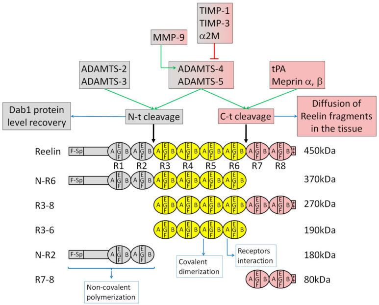
During embryonic development and adulthood, Reelin exerts several important functions in the brain including the regulation of neuronal migration, dendritic growth and branching, dendritic spine formation, synaptogenesis and synaptic plasticity. As a consequence, the Reelin signaling pathway has been associated with several human brain disorders such as lissencephaly, autism, schizophrenia, bipolar disorder, depression, mental retardation, Alzheimer's disease and epilepsy. Several elements of the signaling pathway are known. Core components, such as the Reelin receptors very low-density lipoprotein receptor (VLDLR) and Apolipoprotein E receptor 2 (ApoER2), Src family kinases Src and Fyn, and the intracellular adaptor Disabled-1 (Dab1), are common to most but not all Reelin functions. Other downstream effectors are, on the other hand, more specific to defined tasks. Reelin is a large extracellular protein, and some aspects of the signal are regulated by its processing into smaller fragments. Rather than being inhibitory, the processing at two major sites seems to be fulfilling important physiological functions. In this review, I describe the various cellular events regulated by Reelin and attempt to explain the current knowledge on the mechanisms of action. After discussing the shared and distinct elements of the Reelin signaling pathway involved in neuronal migration, dendritic growth, spine development and synaptic plasticity, I briefly outline the data revealing the importance of Reelin in human brain disorders.

Keywords: Reelin; cellular pathways; cerebral cortex; dendrites; embryonic development; migration; neurodevelopmental disorders; neuron; postnatal maturation; proteolytic processing; signal transduction; synapse.


Conflict of interest statement

The author declares no competing financial interests.

Figures

Figure 1
Figure 1
Structure of Reelin and its processing fragments: Reelin is a large extracellular matrix protein of 450 kDa. The protein starts with a signal peptide followed by an F-spondin homology domain (F-sp) and a unique region. The main body consists of eight Reelin-specific repeats: R1 to R8, each composed by two sub-repeats (A and B) flanking an EGF-like motif (EGF). Reelin ends with a basic stretch of 33 amino acids (++). After its secretion, Reelin is cleaved at two major sites to produce 5 fragments named N-R6, R3-8, R3-6, N-R2 and R7-8. R3-6 is the smallest biologically active fragment. Interaction with Apolipoprotein E receptor 2 (ApoER2) and very low-density lipoprotein receptor (VLDLR) occurs through the first subdomain of R6. Covalent homo-dimerization, which occurs through the first subdomain of R5, is not necessary for receptor interaction but is needed to induce Dab1 phosphorylation. N-R2 is involved in non-covalent polymerization. Proteases involved in the N-terminal (N-t) cleavage between Reelin repeat domains R2 and R3 are shown in grey. Proteases involved in the C-terminal (C-t) cleavage between Reelin repeat domains R6 and R7 are shown in red. Proteases involved in both cleavages are shown in both grey and red. A third cleavage by proprotein convertases removes six amino acids at the C-terminus of Reelin (not shown). The N-terminal cleavage does not prevent the early and midterm events of Reelin signaling but rather might be important to allow Dab1 protein level recovery, preventing an excessive long-term downregulation of the intracellular signal. C-terminal processing is important during the development of the neocortex to allow the active central fragment of Reelin (R3-6) and the N-R6 fragment to diffuse within the tissue, enabling them to reach and trigger the signal in multipolar neurons.
Figure 2
Figure 2
Modeling of the multiple functions of Reelin during migration, dendritic growth and synaptic development and the corresponding signaling pathways: (A,A’). During the early steps of cortical development, early born neurons perform a radial glia-independent somal translocation. Reelin triggers an interaction between Cajal–Retzius cells (CR) and the neuron leading process through N-cadherin and nectins. (B,B’,B’’). As the neocortex develops, the cerebral wall increases in thickness. The somal translocation mode of migration is gradually replaced by a radial migration subdivided into four steps: a short bipolar migration during which postmitotic cells move away from the ventricular zone (VZ) (not depicted here), followed by a radial glia-independent multipolar migration. Cells then switch to a radial glia-dependent but Reelin-independent bipolar locomotion and finish their journey with a terminal somal translocation. While the full-length Reelin is produced and mainly located at the marginal zone (MZ), cleavage fragments of Reelin depleted of the C-t fragment, namely R3-6, N-R2 and N-R6, diffuse from the MZ into the depth of the tissue to reach and signal to multipolar neurons. R3-6 and/or N-R6 interaction with ApoER2 receptors induces the movement of multipolar neurons towards the cortical plate (CP) and a timely subsequent switch from multipolar to bipolar migration. This depends on a Reelin-triggered interaction and activation of N-cadherin with fibroblast growth factors (FGF) receptors (FGFRs) and the downstream effectors Extracellular signal-regulated protein kinases 1 and 2 (Erk1/2). When bipolar locomoting neurons reach the top of the cortical plate (CP), Reelin triggers the detachment from the radial glia and induces a switch from the locomotion mode of migration into the terminal somal translocation. This requires the interaction of full-length Reelin and/or any of its processing fragments containing the central part R3-6 with VLDLR and ApoER2 to stimulate the interaction of Integrins α5β1 on the neuron leading process with fibronectin located at the MZ. Both multipolar migration and terminal somal translocation require activation of the small GTPase Rap1 and of the Phosphoinositide 3-kinase (PI3K)/Akt/n-cofilin pathway. (C,C’). After completion of migration, the leading process transforms into apical dendrites. Reelin triggers dendritic growth and branching in the cerebral cortex and hippocampus. Several mechanisms are involved. All these events seem to be regulated by PI3K: Activation of the mammalian target of rapamycin (mTor)/S6 kinase 1 (S6K1) pathway that could regulate protein synthesis; regulation of the actin cytoskeleton through n-cofilin; and promotion of the translocation of the Golgi into the longest dendrite in a Cdc42-dependent manner. (D,D’). Dendritic spines are small protrusions arising from the dendritic shaft where most excitatory synapses reside. Reelin regulates their density in the hippocampus and the cerebral cortex. Synaptic plasticity is the ability of a synapse to modify its strength and is also influenced by Reelin that accumulates at synaptic contacts. N-methyl-D-aspartate (NMDA) and α-amino-3-hydroxy-5-methyl-4-isoxazolepropionic acid (AMPA)-type glutamate receptors (NMDARs and AMPARs ) are two postsynaptic ionotropic receptors directly involved in synaptic plasticity. Reelin modulates the subunit composition of NMDARs, its phosphorylation, its calcium conductance and potentiation. Reelin also facilitates the insertion of AMPARs in postsynaptic membranes and presynaptic release of neurotransmitters. Core components VLDLR and/or ApoER2, Src family kinases (SFK) and Dab1 are involved in most Reelin functions. Dab1 is polyubiquitinated by an E3 ubiquitin ligase complex containing Cullin 5 (Cul5), Rbx2 and SOCS7 and is subsequently degraded by the proteasome. This rapid downregulation of the signal is necessary for a correct organization of the CP. Its function in Reelin-induced dendritic growth and at the synapse has not been investigated. The PI3K/Akt pathway is also involved in all these Reelin functions but was apparently not crucial for early somal translocation. See the text for more details.

Similar articles

Cited by

References

    1. Falconer D.S. Two new mutants, ‘trembler’ and ‘reeler’, with neurological actions in the house mouse (Mus musculus L.) J. Genet. 1951;50:192–205. doi: 10.1007/BF02996215. - DOI - PubMed
    1. Caviness V.S., So D.K., Sidman R.L. The Hybrid Reeler Mouse. J. Hered. 1972;63:241–246. doi: 10.1093/oxfordjournals.jhered.a108286. - DOI - PubMed
    1. Hamburgh M. Analysis of the postnatal developmental effects of “reeler,” a neurological mutation in mice. A study in developmental genetics. Dev. Biol. 1963;8:165–185. doi: 10.1016/0012-1606(63)90040-X. - DOI - PubMed
    1. De Rouvroit C.L., Goffinet A.M. Factors Influencing Mammalian Kidney Development: Implications for Health in Adult Life. Volume 150. Springer Science and Business Media; Berlin/Heidelberg, Germany: 1998. The Reeler Mouse as a Model of Brain Development; pp. 1–106. - DOI
    1. Rice D.S., Nusinowitz S., Azimi A.M., Martínez A., Soriano E., Curran T. The Reelin Pathway Modulates the Structure and Function of Retinal Synaptic Circuitry. Neuron. 2001;31:929–941. doi: 10.1016/S0896-6273(01)00436-6. - DOI - PubMed

Publication types

MeSH terms