. 2020 Jun 26;10(6):1246.
doi: 10.3390/nano10061246.

Fast and Accurate Pneumocystis Pneumonia Diagnosis in Human Samples Using a Label-Free Plasmonic Biosensor

Affiliations

Fast and Accurate Pneumocystis Pneumonia Diagnosis in Human Samples Using a Label-Free Plasmonic Biosensor

Olalla Calvo-Lozano et al. Nanomaterials (Basel). .

Abstract

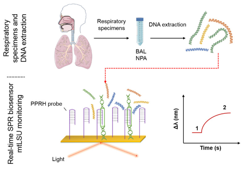
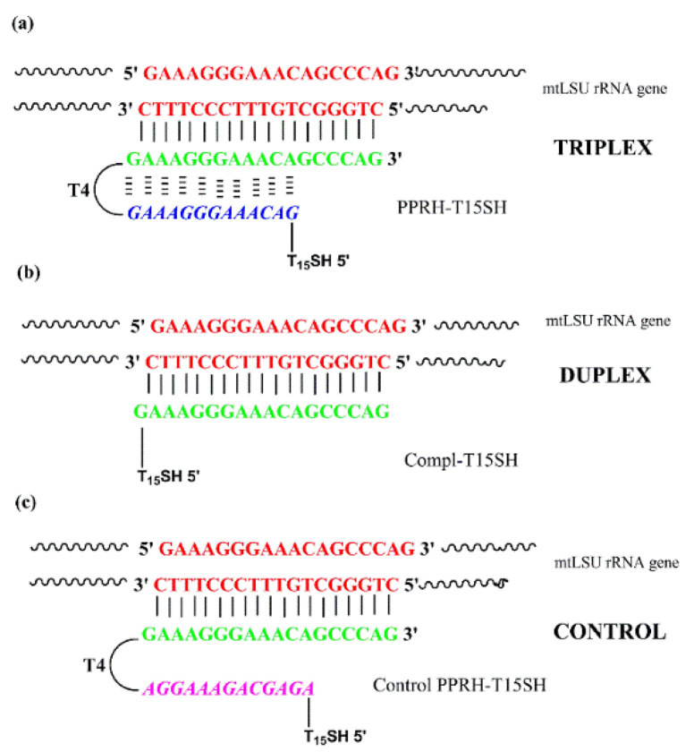
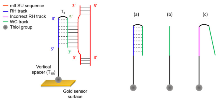
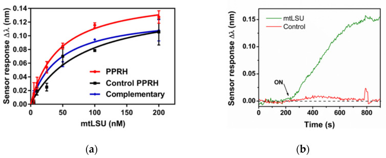
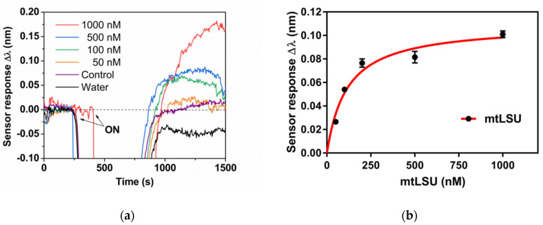
Pneumocystis jirovecii is a fungus responsible for human Pneumocystis pneumonia, one of the most severe infections encountered in immunodepressed individuals. The diagnosis of Pneumocystis pneumonia continues to be challenging due to the absence of specific symptoms in infected patients. Moreover, the standard diagnostic method employed for its diagnosis involves mainly PCR-based techniques, which besides being highly specific and sensitive, require specialized personnel and equipment and are time-consuming. Our aim is to demonstrate an optical biosensor methodology based on surface plasmon resonance to perform such diagnostics in an efficient and decentralized scheme. The biosensor methodology employs poly-purine reverse-Hoogsteen hairpin probes for the detection of the mitochondrial large subunit ribosomal RNA (mtLSU rRNA) gene, related to P. jirovecii detection. The biosensor device performs a real-time and label-free identification of the mtLSU rRNA gene with excellent selectivity and reproducibility, achieving limits of detection of around 2.11 nM. A preliminary evaluation of clinical samples showed rapid, label-free and specific identification of P. jirovecii in human lung fluids such as bronchoalveolar lavages or nasopharyngeal aspirates. These results offer a door for the future deployment of a sensitive diagnostic tool for fast, direct and selective detection of Pneumocystis pneumonia disease.

Keywords: DNA capture; Pneumocystis jirovecii; Surface Plasmon Resonance; clinical diagnosis; optical biosensor; triplex.


Conflict of interest statement

The authors declare no conflicts of interest.

Figures

Figure A1
Figure A1
Custom-designed SPR biosensor. (a) Photograph of the experimental SPR set-up. (b) Scheme of the sensing principle of a SPR biosensor. (c) Graph showing the displacement of the resonance peak (∆λ) to RI changes (photon vs. λ (nm)). Real-time monitoring of wavelength displacements (Δλ vs. time (s)).
Figure A2
Figure A2
Optimization of the methodology for selective detection of the mtLSU rRNA gene. (a) Detection signals obtained by employing different surface SAMs in the immobilization of PPRH probes. (b) Effect of changes in the ionic strength of the hybridization buffer on the mtLSU rRNA recognition using PPRH probes. (c) Formamide effect in the cross-hybridization response of PPRH probes.
Figure A3
Figure A3
CD spectra of duplex (Compl + ss-DNA mtLSU target) and antiparallel triplex (PPRH + ss-DNA mtLSU target).
Figure 1
Figure 1
Surface plasmon resonance (SPR) biosensor methodology for the specific detection of P. jirovecii. The samples (BAL and NPA) after DNA extraction were injected to the SPR device in which the PPRH probe is attached above the gold surface of the sensor. Wavelength variation of the reflected light is directly related to the amount of the analyte bounded to the bioreceptor. (BAL: bronchoalveolar lavage, NPA: nasopharyngeal aspirates, PPRH: polypurine reverse-Hoogsteen hairpin, 1: Wavelength correspond to the baseline, 2: Wavelength correspond to target analyte recognition).
Figure 2
Figure 2
Sequences designed for use as bioreceptors in the SPR biosensor for the detection of the mtLSU rRNA gene of Pneumocystis jirovecii. (a) PPRH-T15SH capture probe recognizes the pyrimidine sequence of the mtLSU rRNA gene and forms a stable antiparallel triplex structure. (b) Complementary–T15SH forms a duplex structure with the pyrimidine sequence of the mtLSU rRNA. (c) Control PPHR-T15SH forms a duplex with the pyrimidine sequence of the mtLSU rRNA gene.
Figure 3
Figure 3
Schematic representation of the mtLSU rRNA gene capture using the PPRH probe, forming an antiparallel triplex structure. In addition, different bioreceptors employed in the SPR biosensor are shown: (a) PPRH probe, (b) complementary probe, (c) control PPRH probe. (Colors correspond to Figure 2).
Figure 4
Figure 4
mtLSU rRNA gene detection. (a) Calibration curves of mtLSU rRNA on SSC 2.5 X + 5% FA buffer. Sensor response represents the mean ± SD of three measurements in a PPRH: CH3–PEG (1:1) receptor monolayer. (b) Real-time monitoring of the wavelength displacements (Δλ vs. time) corresponding to the hybridization of 100 nM mtLSU rRNA analyte and DNA control sequences using SSC 2.5 X + 5% FA buffer.
Figure 5
Figure 5
mtLSU rRNA detection. (a) Real-time monitoring of wavelength displacements (Δλ vs time) corresponding to the hybridization of different mtLSU rRNA concentrations (1000, 500, 100 and 50 nM, respectively) diluted in water, control DNA and water in a PPRH: CH3–PEG–SH (1:1) monolayer. (b) Calibration curves of mtLSU rRNA on water using SSC 2.5 X + 5% FA as running buffer. Sensor response represents the mean ± SD of three measurements.
Figure 6
Figure 6
Detection of mtLSU rRNA gene contained in synthetic pGEM-T Easy plasmids and DNA fragments. (a) Sensor signal corresponding to pGEM-T Easy plasmids (P) and ds-DNA fragments (F). (b) Sensor signal corresponding to pGEM-T Easy plasmids (P) and pGEM-T Easy plasmids digested (D) by EcoRI enzyme. For all the measurements SSC 2.5 X + 5% FA as running buffer and PPRH: CH3–PEG (1:1) monolayers were performed.
Figure 7
Figure 7
Analysis of mtLSU rRNA in clinical samples from patients infected by (i) Pseudomones (n = 4), (ii) Cladosporium (n = 4) and (iii) Pneumocystis (n = 4), performed using the SPR biosensor biofunctionalized with PPRH: CH3–PEG (1:1) monolayer. Representation of one-way ANOVA test where median, maximum and minimum values are shown. One-way ANOVA test, p-value = 0.0258.

Similar articles

Cited by

References

    1. Pereira-Díaz E., Moreno-Verdejo F., de la Horra C., Guerrero J.A., Calderón E.J., Medrano F.J. Changing Trends in the Epidemiology and Risk Factors of Pneumocystis Pneumonia in Spain. Front. Public Health. 2019;7:275. doi: 10.3389/fpubh.2019.00275. - DOI - PMC - PubMed
    1. Morris A., Norris K.A. Colonization by pneumocystis jirovecii and its role in disease. Clin. Microbiol. Rev. 2012;25:297–317. doi: 10.1128/CMR.00013-12. - DOI - PMC - PubMed
    1. Wang H.W., Lin C.C., Kuo C.F., Liu C.P., Lee C.M. Mortality predictors of Pneumocystis jirovecii pneumonia in human immunodeficiency virus-infected patients at presentation: Experience in a tertiary care hospital of northern Taiwan. J. Microbiol. Immunol. Infect. 2011;44:274–281. doi: 10.1016/j.jmii.2010.08.006. - DOI - PubMed
    1. Calderón E.J. Pneumocystis Infection: Seeing beyond the Tip of the Iceberg. Clin. Infect. Dis. 2010;50:354–356. doi: 10.1086/649870. - DOI - PubMed
    1. Tasaka S. Recent Advances in the Diagnosis and Management of Pneumocystis Pneumonia. Tuberc. Respir. Dis. 2020;83:132–140. doi: 10.4046/trd.2020.0015. - DOI - PMC - PubMed