Review
. 2020 Jun 27;12(7):693.
doi: 10.3390/v12070693.

Viral Membrane Fusion and the Transmembrane Domain

Affiliations
Review

Viral Membrane Fusion and the Transmembrane Domain

Chelsea T Barrett et al. Viruses. .

Abstract

Initiation of host cell infection by an enveloped virus requires a viral-to-host cell membrane fusion event. This event is mediated by at least one viral transmembrane glycoprotein, termed the fusion protein, which is a key therapeutic target. Viral fusion proteins have been studied for decades, and numerous critical insights into their function have been elucidated. However, the transmembrane region remains one of the most poorly understood facets of these proteins. In the past ten years, the field has made significant advances in understanding the role of the membrane-spanning region of viral fusion proteins. We summarize developments made in the past decade that have contributed to the understanding of the transmembrane region of viral fusion proteins, highlighting not only their critical role in the membrane fusion process, but further demonstrating their involvement in several aspects of the viral lifecycle.

Keywords: transmembrane domain; viral fusion protein.


Conflict of interest statement

The authors report no conflict of interest.

Figures
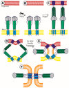
Figure 1
Figure 1
A model of viral membrane fusion function. This figure depicts a model of fusion mediated by a Class I viral fusion protein; however, related processes occur in the case of cClass II and III viral fusion proteins as well. (A) The fusion protein situates itself in the viral membrane (yellow). The first step of viral fusion is a priming event; in the case of Class I proteins, the protein itself undergoes the proteolytic processing to prime it for fusion. For Class II fusion proteins, it is a companion protein that gets proteolytically processed; (B) Once primed, the viral fusion protein remains in a metastable, pre-fusion conformation until it receives a triggering signal; (C) Upon receipt of the triggering signal, the protein extends out, forming a pre-hairpin structure, allowing for the fusion peptide or fusion loop (dark blue) to enter the target membrane (red); (D) This extended structure then begins to fold back on itself, bringing the N-terminal and C-terminal heptad repeats closer (dark green and light green, respectively), and in turn pulling the viral membrane and target membrane together; (E) As the N-terminal and C terminal heptad repeats zipper together to form a six-helix bundle, the target and viral membrane reach a hemi-fusion state, in which the outer leaflets have started to mix (orange); (F) Finally, the fusion peptide and transmembrane domain (light blue) come into close proximity to complete the merging of the two membranes and opening of the fusion pore. This final structure of a trimer of hairpins is a common conformation among all viral fusion proteins [9].
Figure 2
Figure 2
Full-length structures of viral fusion proteins. While the ectodomain structures of numerous viral fusion proteins have been solved, only a few solved structures of full-length viral fusion proteins, including the transmembrane domain (TMD) have been solved. (A) Full-length influenza hemagglutinin (HA) was solved in 2018 [51]. Two structures were published, 6HJQ (far left) and 6HQR (far right). The first is HA in its pre-fusion conformation, and the ectodomain is in line with the TMD helices (middle left, zoomed in). In the second structure, the ectodomain is tilted 52° with respect to the TMD helices (middle right, zoomed in); this likely represents a scenario that is an intermediate state of fusion. In each zoomed-in area, a linker region is indicated (slate), and this region remains flexible to help compensate for this tilt. (B) In 2013, the structure of full-length Dengue virus E protein was solved [53]. This structure showed the hetero-tetramer complex of two E proteins (light and dark purple) and 2 M proteins (gray) with their respective TMD helices (3J2P). Flavivirus E proteins have two TMDs that have extensive hydrophobic interactions between them. The structure also includes three peri-membrane helices that lie on the outer surface of the viral membrane, approximately perpendicular to the TMD helices. (C) Full-length Herpes simplex virus 1 gB protein was published in 2018 [54]. This structure (5V2S) shows the three TMD helices situated in a triangular teepee structure; the MPER (dark grey) is a helix that lies almost perpendicular to the orientation of the TMD helices. The solved structure also includes a large portion of the cytoplasmic tail (CTD, light gray). Each CTD has two helices, the first of which is a small helix that links to a larger helix which then angles back towards the inner leaflet of the viral membrane. Because of the orientation, these CTDs may act as a clamp that assists in holding the gB TMDs in specific conformation, and the angle of the CTD may work in concert with the TMD helices to dictate the overall conformation of the protein. Figure made with PyMOL (Schrödinger®, Neo York, NY, USA).

Similar articles

Cited by

References

    1. Earp L.J., Delos S.E., Park H.E., White J.M. The many mechanisms of viral membrane fusion proteins. Curr. Top. Microbiol. Immunol. 2005;285:25–66. doi: 10.1007/3-540-26764-6_2. - DOI - PMC - PubMed
    1. Harrison S.C. Viral membrane fusion. Nat. Struct. Mol. Biol. 2008;15:690–698. doi: 10.1038/nsmb.1456. - DOI - PMC - PubMed
    1. Harrison S.C. Viral membrane fusion. Virology. 2015;479–480:498–507. doi: 10.1016/j.virol.2015.03.043. - DOI - PMC - PubMed
    1. Dutch R.E., Jardetzky T.S., Lamb R.A. Virus membrane fusion proteins: Biological machines that undergo a metamorphosis. Biosci. Rep. 2000;20:597–612. doi: 10.1023/A:1010467106305. - DOI - PubMed
    1. White J.M., Whittaker G.R. Fusion of Enveloped Viruses in Endosomes. Traffic. 2016;17:593–614. doi: 10.1111/tra.12389. - DOI - PMC - PubMed

Publication types

Substances

LinkOut - more resources