. 2020 Jun 27;11(7):628.
doi: 10.3390/mi11070628.

Analysis of Single Nucleotide-Mutated Single-Cancer Cells Using the Combined Technologies of Single-Cell Microarray Chips and Peptide Nucleic Acid-DNA Probes

Affiliations

Analysis of Single Nucleotide-Mutated Single-Cancer Cells Using the Combined Technologies of Single-Cell Microarray Chips and Peptide Nucleic Acid-DNA Probes

Hajime Shigeto et al. Micromachines (Basel). .

Abstract

Research into cancer cells that harbor gene mutations relating to anticancer drug-resistance at the single-cell level has focused on the diagnosis of, or treatment for, cancer. Several methods have been reported for detecting gene-mutated cells within a large number of non-mutated cells; however, target single nucleotide-mutated cells within a large number of cell samples, such as cancer tissue, are still difficult to analyze. In this study, a new system is developed to detect and isolate single-cancer cells expressing the T790M-mutated epidermal growth factor receptor (EGFR) mRNA from multiple non-mutated cancer cells by combining single-cell microarray chips and peptide nucleic acid (PNA)-DNA probes. The single-cell microarray chip is made of polystyrene with 62,410 microchambers (31-40 µm diameter). The T790M-mutated lung cancer cell line, NCI-H1975, and non-mutated lung cancer cell line, A549, were successfully separated into single cells in each microchambers on the chip. Only NCI-H1975 cell was stained on the chip with a fluorescein isothiocyanate (FITC)-conjugated PNA probe for specifically detecting T790M mutation. Of the NCI-H1975 cells that spiked into A549 cells, 0-20% were quantitatively analyzed within 1 h, depending on the spike concentration. Therefore, our system could be useful in analyzing cancer tissue that contains a few anticancer drug-resistant cells.

Keywords: T790M mutation; cell microarray; epidermal growth factor receptor (EGFR); lung cancer; peptide nucleic acid (PNA) probe; single nucleotide mutation; single-cell analysis.


Conflict of interest statement

The authors declare no competing financial interests.

Figures
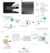
Figure 1
Figure 1
Schematic concept for the analysis of single nucleotide-mutated cancer cells. (a) Photograph of a real single-cell microarray chip device, which was made with polystyrene and consists of 62,410 microchambers (upper diameter: 31–40 µm, lower diameter: 11–20 µm, depth: 28 µm, pitch: 100 µm). Each single-cell microarray chip consisted of 10 different-sized patterns of clusters, and each cluster consisted of 6241 (79 × 79) microchambers. Each microchamber had the shape of a circular cone frustum. (b) Microscopic image of microchambers (upper diameter: 32 µm, lower diameter: 12 µm) of one cluster in the single-cell microarray chip. The picture shows single lung cancer cells occupying each microchamber. (c) Principle of target single nucleotide-mutated epidermal growth factor receptor (EGFR) mRNA detection with a peptide nucleic acid (PNA)-DNA probe. (d) Schematic process for the analysis of mutation-harboring cancer cells at the single-cell level using our original techniques combined with a single-cell microarray chip and a PNA-DNA probe.
Figure 2
Figure 2
Separation and analysis of different lung cancer cell lines (NCI-H1975 and A549) on the single-cell microarray chip. The upper and lower figures show the representative fluorescent images of NCI-H1975 (T790M-mutated) and A549 (non-mutated) cells, respectively, stained by diluted 4,6-diamidino-2-phenylindole (DAPI; blue), fluorescein isothiocyanate (FITC)-conjugated peptide nucleic acid (PNA)-DNA probes (green), and phycoerythrin (PE)-labeled cytokeratin (CK) antibody (red).
Figure 3
Figure 3
Analysis of the ratio of T790M-mutated cancer cells (NCI-H1975) spiked into non-mutated cells (A549) using combination technology with the single-cell microarray chip and peptide nucleic acid (PNA)-DNA probe. (a) Representative fluorescent images of 5%, 10%, and 20% NCI-H1975 cells spiking into A549 cells stained with diluted 4,6-diamidino-2-phenylindole (DAPI; blue), fluorescein isothiocyanate (FITC)-conjugated PNA-DNA probes (green), and phycoerythrin (PE)-labeled cytokeratin (CK) antibody (red). White arrows indicate positive cells (T790M-mutated). (b) Information on the number and ratio of T790M-mutated cells (NCI-H1975) spiked into A549 cells. (c) Linearity plots for the spiked and detected ratio of T790M-mutated cells (NCI-H1975) spiked into A549 cells. R2 indicates the correlation coefficient. The results are shown as the mean ± standard error of the three replicates.

Similar articles

Cited by

References

    1. Di Carlo D., Lee L.P. Dynamic single-cell analysis for quantitative biology. Anal. Chem. 2006;78:7918–7925. doi: 10.1021/ac069490p. - DOI - PubMed
    1. Shigeto H., Yamamura S. Bioluminescence resonance energy transfer (BRET)-based biosensing probes using novel luminescent and fluorescent protein pairs. Sens. Mater. 2019;31:71–78. doi: 10.18494/SAM.2019.2049. - DOI
    1. Shigeto H., Ono T., Ikeda T., Hirota R., Ishida T., Kuroda A., Funabashi H. Insulin sensor cells for the analysis of insulin secretion responses in single living pancreatic β cells. Analyst. 2019;144:3765–3772. doi: 10.1039/C9AN00405J. - DOI - PubMed
    1. Yamamura S., Kishi H., Tokimitsu Y., Kondo S., Honda R., Ramachandra Rao S., Omori M., Tamiya E., Muraguchi A. Single-cell microarray for analyzing cellular response. Anal. Chem. 2005;77:8050–8056. doi: 10.1021/ac0515632. - DOI - PubMed
    1. Yatsushiro S., Yamamura S., Yamaguchi Y., Shinohara Y., Tamiya E., Horii T., Baba Y., Kataoka M. Rapid and highly sensitive detection of malaria-infected erythrocytes using a cell microarray chip. PLoS ONE. 2010;5:e13179. doi: 10.1371/journal.pone.0013179. - DOI - PMC - PubMed