

A Pilot Study of Musculoskeletal Abnormalities in Patients in Recovery from a Unilateral Rupture-Repaired Achilles Tendon
- PMID: 32605170
- PMCID: PMC7369810
- DOI: 10.3390/ijerph17134642
A Pilot Study of Musculoskeletal Abnormalities in Patients in Recovery from a Unilateral Rupture-Repaired Achilles Tendon
Abstract
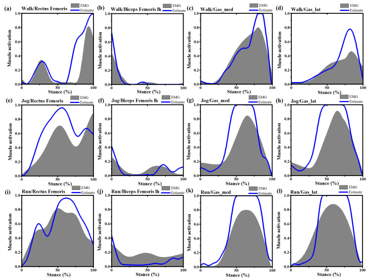
The purpose of this study was to compare the inter-limb joint kinematics, joint moments, muscle forces, and joint reaction forces in patients after an Achilles tendon rupture (ATR) via subject-specific musculoskeletal modeling. Six patients recovering from a surgically repaired unilateral ATR were included in this study. The bilateral Achilles tendon (AT) lengths were evaluated using ultrasound imaging. The three-dimensional marker trajectories, ground reaction forces, and surface electromyography (sEMG) were collected on both sides during self-selected speed during walking, jogging and running. Subject-specific musculoskeletal models were developed to compute joint kinematics, joint moments, muscle forces and joint reaction forces. AT lengths were significantly longer in the involved side. The side-to-side triceps surae muscle strength deficits were combined with decreased plantarflexion angles and moments in the injured leg during walking, jogging and running. However, the increased knee extensor femur muscle forces were associated with greater knee extension degrees and moments in the involved limb during all tasks. Greater knee joint moments and joint reaction forces versus decreased ankle joint moments and joint reaction forces in the involved side indicate elevated knee joint loads compared with reduced ankle joint loads that are present during normal activities after an ATR. In the frontal plane, increased subtalar eversion angles and eversion moments in the involved side were demonstrated only during jogging and running, which were regarded as an indicator for greater medial knee joint loading. It seems after an ATR, the elongated AT accompanied by decreased plantarflexion degrees and calf muscle strength deficits indicates ankle joint function impairment in the injured leg. In addition, increased knee extensor muscle strength and knee joint loads may be a possible compensatory mechanism for decreased ankle function. These data suggest patients after an ATR may suffer from increased knee overuse injury risk.
Keywords: Achilles tendon rupture; ankle; gait; knee; musculoskeletal modeling.
Conflict of interest statement
The authors declare no conflicts of interest.
Figures


References
-
- Ganestam A., Kallemose T., Troelsen A., Barfod K.W. Increasing incidence of acute Achilles tendon rupture and a noticeable decline in surgical treatment from 1994 to 2013. A nationwide registry study of 33,160 patients. Knee Surg. Sports Traumatol. Arthrosc. 2016;24:3730–3737. doi: 10.1007/s00167-015-3544-5. - DOI - PubMed
-
- Barfod K.W., Bencke J., Lauridsen H.B., Dippmann C., Ebskov L., Troelsen A. Nonoperative, dynamic treatment of acute achilles tendon rupture: Influence of early weightbearing on biomechanical properties of the plantar flexor muscle-tendon complex-a blinded, randomized, controlled trial. J. Foot Ankle Surg. 2015;54:220–226. doi: 10.1053/j.jfas.2014.11.018. - DOI - PubMed
-
- Heikkinen J., Lantto I., Flinkkila T., Ohtonen P., Niinimaki J., Siira P., Laine V., Leppilahti J. Soleus Atrophy is Common after the Nonsurgical Treatment of Acute Achilles Tendon Ruptures: A Randomized Clinical Trial Comparing Surgical and Nonsurgical Functional Treatments. Am. J. Sports Med. 2017;45:1395–1404. doi: 10.1177/0363546517694610. - DOI - PubMed
Publication types
MeSH terms
LinkOut - more resources
Full Text Sources
Miscellaneous
