

Androgens' effects on cerebrovascular function in health and disease
- PMID: 32605602
- PMCID: PMC7328272
- DOI: 10.1186/s13293-020-00309-4
Androgens' effects on cerebrovascular function in health and disease
Abstract
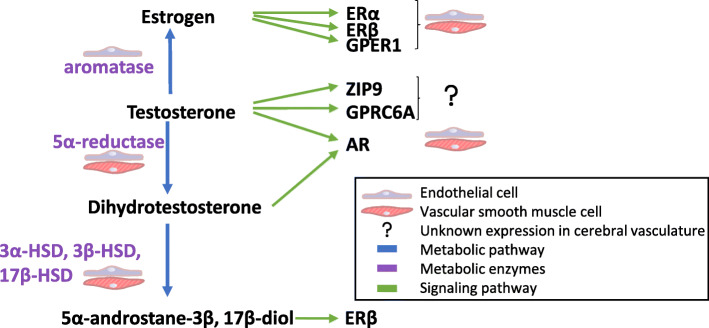
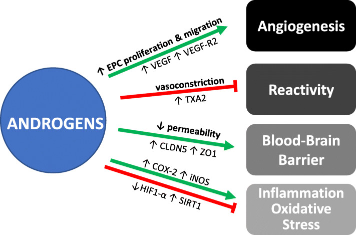
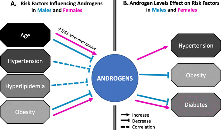
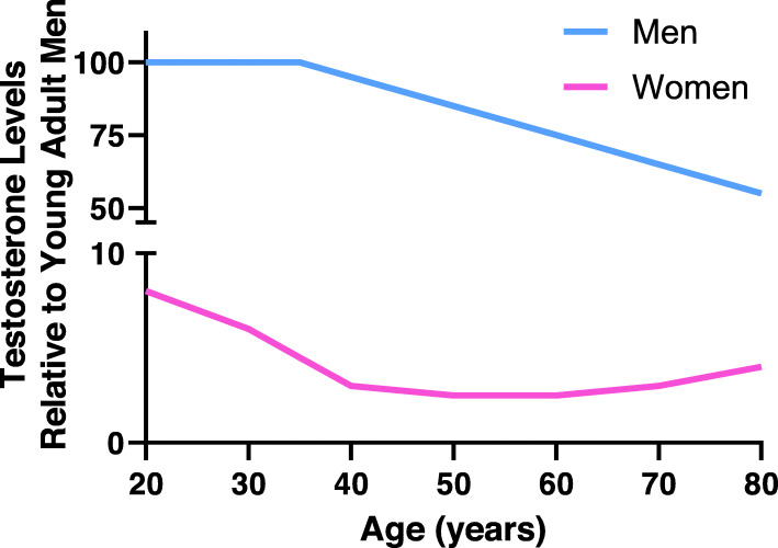
Androgens affect the cerebral vasculature and may contribute to sex differences in cerebrovascular diseases. Men are at a greater risk for stroke and vascular contributions to cognitive impairment and dementia (VCID) compared to women throughout much of the lifespan. The cerebral vasculature is a target for direct androgen actions, as it expresses several sex steroid receptors and metabolizing enzymes. Androgens' actions on the cerebral vasculature are complex, as they have been shown to have both protective and detrimental effects, depending on factors such as age, dose, and disease state. When administered chronically, androgens are shown to be pro-angiogenic, promote vasoconstriction, and influence blood-brain barrier permeability. In addition to these direct effects of androgens on the cerebral vasculature, androgens also influence other vascular risk factors that may contribute to sex differences in cerebrovascular diseases. In men, low androgen levels have been linked to metabolic and cardiovascular diseases including hypertension, diabetes, hyperlipidemia, and obesity, which greatly increase the risk of stroke and VCID. Thus, a better understanding of androgens' interactions with the cerebral vasculature under physiological and pathological conditions is of key importance.
Keywords: Androgens; Blood-brain barrier; Cerebral vasculature; Cerebrovascular disease; Dihydrotestosterone; Endothelial; Hormone therapy; Stroke; Testosterone; Vascular contributions to cognitive impairment and dementia.
Conflict of interest statement
The authors declare that they have no competing interests.
Figures
References
-
- Armulik A, Genove G, Mae M, Nisancioglu MH, Wallgard E, Niaudet C, et al. Pericytes regulate the blood-brain barrier. Nature. 2010;468(7323):557–561. - PubMed
-
- Quaegebeur A, Lange C, Carmeliet P. The neurovascular link in health and disease: molecular mechanisms and therapeutic implications. Neuron. 2011;71(3):406–424. - PubMed
-
- Dyrna F, Hanske S, Krueger M, Bechmann I. The blood-brain barrier. J NeuroImmune Pharmacol. 2013;8(4):763–773. - PubMed
Publication types
MeSH terms
Substances
Grants and funding
LinkOut - more resources
Full Text Sources
