

Changes in retinal layer thickness with maturation in the dog: an in vivo spectral domain - optical coherence tomography imaging study
- PMID: 32605619
- PMCID: PMC7329457
- DOI: 10.1186/s12917-020-02390-8
Changes in retinal layer thickness with maturation in the dog: an in vivo spectral domain - optical coherence tomography imaging study
Abstract
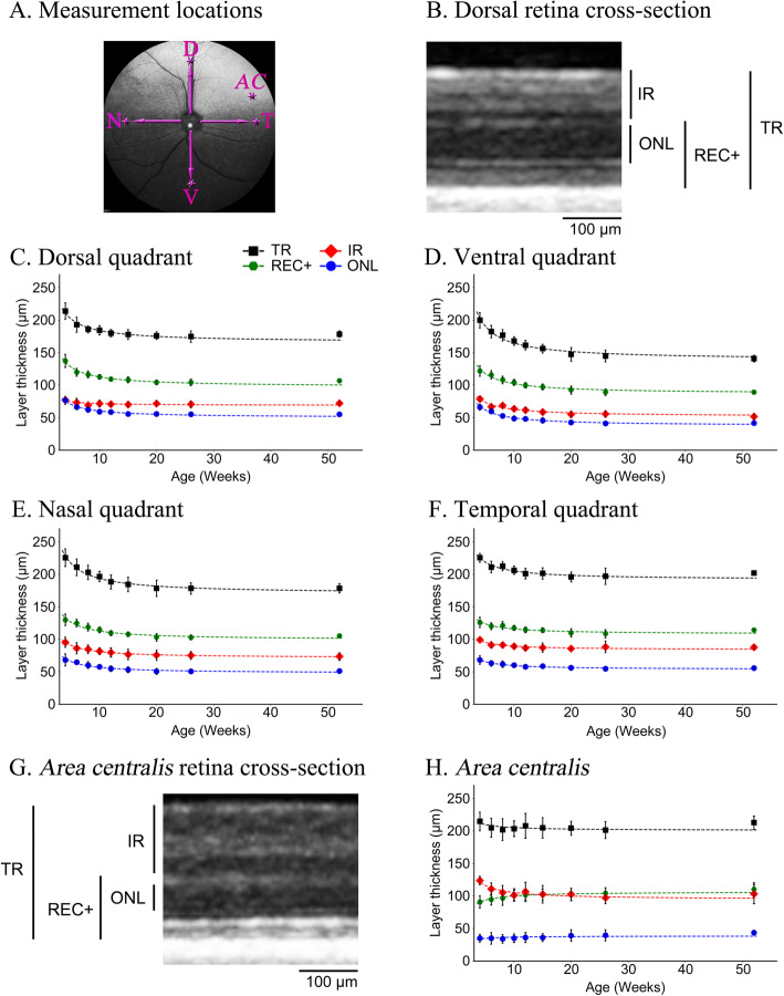
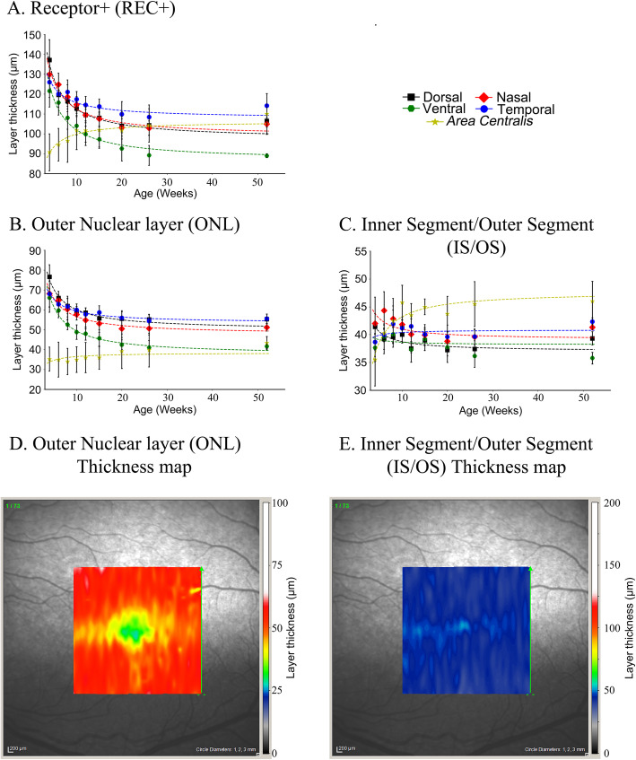
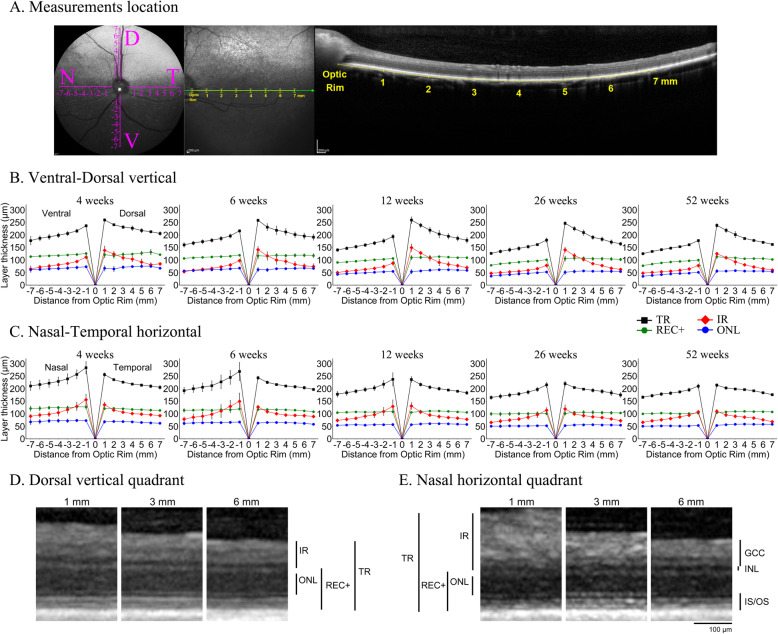
Background: Retinal diseases are common in dogs. Some hereditary retinal dystrophies in dogs are important not only because they lead to vision loss but also because they show strong similarities to the orthologous human conditions. Advances in in vivo non-invasive retinal imaging allow the capture of retinal cross-section images that parallel low power microscopic examination of histological sections. Spectral domain - optical coherence tomography (SD-OCT) allows the measurement of retinal layer thicknesses and gives the opportunity for repeat examination to investigate changes in thicknesses in health (such as changes with maturation and age) and disease (following the course of retinal degenerative conditions). The purpose of this study was to use SD-OCT to measure retinal layer thicknesses in the dog during retinal maturation and over the first year of life. SD-OCT was performed on normal beagle cross dogs from 4 weeks of age to 52 weeks of age. To assess changes in layer thickness with age, measurements were taken from fixed regions in each of the 4 quadrants and the area centralis (the region important for most detailed vision). Additionally, changes in retinal layer thickness along vertical and horizontal planes passing through the optic nerve head were assessed.
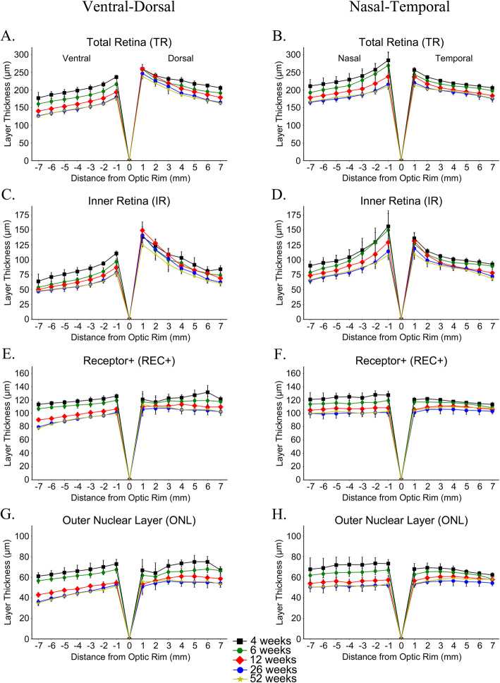
Results: In the four quadrants an initial thinning of retinal layers occurred over the first 12 to 15 weeks of life after which there was little change in thickness. However, in the area centralis there was a thickening of the photoreceptor layer over this time period which was mostly due to a lengthening of the photoreceptor inner/outer segment layer. The retina thinned with greater distances from the optic nerve head in both vertical and horizontal planes with the dorsal retina being thicker than the ventral retina. Most of the change in thickness with distance from the optic nerve head was due to difference in thickness of the inner retinal layers. The outer retinal layers remained more constant in thickness, particularly in the horizontal plane and dorsal to the optic nerve head.
Conclusions: These measurements will provide normative data for future studies.
Keywords: Area centralis; Dog; Maturation; Retina; SD-OCT.
Conflict of interest statement
Not applicable.
Figures
Similar articles
-
Outer retinal thickness and visibility of the choriocapillaris in four distinct retinal regions imaged with spectral domain optical coherence tomography in dogs and cats.Vet Ophthalmol. 2022 May;25 Suppl 1(Suppl 1):122-135. doi: 10.1111/vop.12989. Epub 2022 May 25. Vet Ophthalmol. 2022. PMID: 35611616 Free PMC article.
-
Evaluation of retinal morphology of canine sudden acquired retinal degeneration syndrome using optical coherence tomography and fluorescein angiography.Vet Ophthalmol. 2019 Jul;22(4):398-406. doi: 10.1111/vop.12602. Epub 2018 Aug 22. Vet Ophthalmol. 2019. PMID: 30136357
-
Comparison of chorioretinal layers in rhesus macaques using spectral-domain optical coherence tomography and high-resolution histological sections.Exp Eye Res. 2018 Mar;168:69-76. doi: 10.1016/j.exer.2018.01.012. Epub 2018 Jan 17. Exp Eye Res. 2018. PMID: 29352993 Free PMC article.
-
Spectral domain optical coherence tomography (SD-OCT) assessment of the healthy female canine retina and optic nerve.Vet Ophthalmol. 2011 Nov;14(6):400-5. doi: 10.1111/j.1463-5224.2011.00887.x. Epub 2011 Apr 19. Vet Ophthalmol. 2011. PMID: 22050777 Review.
-
Central retina changes in Parkinson's disease: a systematic review and meta-analysis.J Neurol. 2021 Dec;268(12):4646-4654. doi: 10.1007/s00415-020-10304-9. Epub 2020 Nov 10. J Neurol. 2021. PMID: 33174132
Cited by
-
Outer retinal thickness and visibility of the choriocapillaris in four distinct retinal regions imaged with spectral domain optical coherence tomography in dogs and cats.Vet Ophthalmol. 2022 May;25 Suppl 1(Suppl 1):122-135. doi: 10.1111/vop.12989. Epub 2022 May 25. Vet Ophthalmol. 2022. PMID: 35611616 Free PMC article.
-
Interspecies Retinal Diversity and Optic Nerve Anatomy in Odontocetes.Animals (Basel). 2023 Nov 6;13(21):3430. doi: 10.3390/ani13213430. Animals (Basel). 2023. PMID: 37958185 Free PMC article.
-
Morphological and Morphometric Analysis of Canine Choroidal Layers Using Spectral Domain Optical Coherence Tomography.Int J Environ Res Public Health. 2023 Feb 10;20(4):3121. doi: 10.3390/ijerph20043121. Int J Environ Res Public Health. 2023. PMID: 36833819 Free PMC article.
-
An unusual inherited electroretinogram feature with an exaggerated negative component in dogs.Vet Ophthalmol. 2022 Sep;25(5):385-397. doi: 10.1111/vop.12998. Epub 2022 Jun 17. Vet Ophthalmol. 2022. PMID: 35713167 Free PMC article.
References
-
- Gum GG, Gelatt KC, Samuelson DA. Maturation of the retina of the canine neonate as determined by electroretinography and histology. Am J of Vet Res. 1984;45(6):1166–1171. - PubMed
-
- Aguirre GD, Rubin LF, Bistner SI. The development of the canine eye. Am J Vet Res. 1972;33(12):2399–2414. - PubMed
-
- Miller WM, Albert RA, Bosinger TR, Holloway CL, Simpson ST, Tojvio-Kinnucan MA. Postnatal development of the photoreceptor inner segment of the retina in dogs. Am J of Vet Res. 1989;50(12):2089–2092. - PubMed
-
- Hassenstein A, Meyer CH. Clinical use and research applications of Heidelberg retinal angiography and spectral-domain optical coherence tomography - a review. Clin Exp Ophthalmol. 2009;37(1):130–143. - PubMed
MeSH terms
Grants and funding
LinkOut - more resources
Full Text Sources
