. 2020 Jul 1;33(3):275-283.
doi: 10.3344/kjp.2020.33.3.275.

Diffusion tensor imaging of the C1-C3 dorsal root ganglia and greater occipital nerve for cervicogenic headache

Affiliations

Diffusion tensor imaging of the C1-C3 dorsal root ganglia and greater occipital nerve for cervicogenic headache

Lang Wang et al. Korean J Pain. .

Abstract

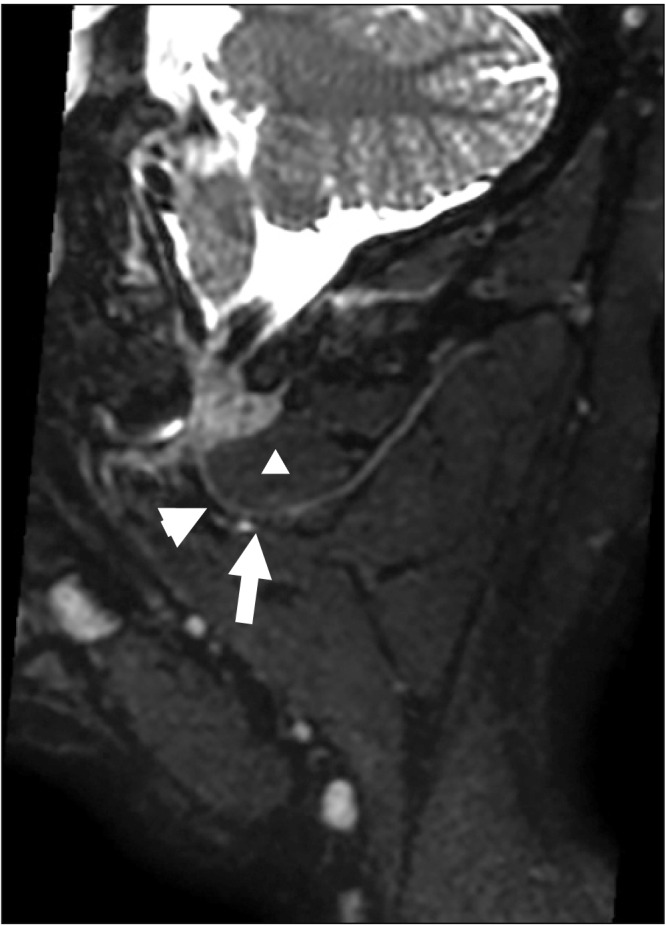
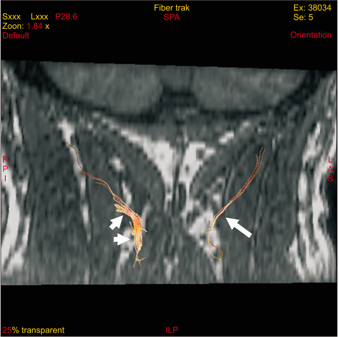
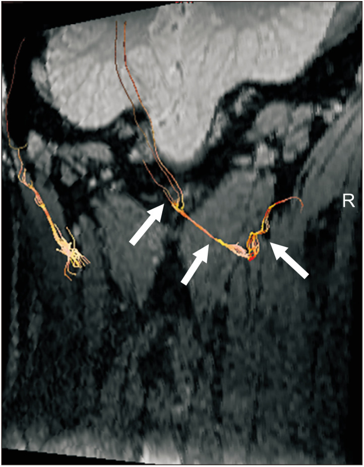
Background: Previous studies showed neurography and tractography of the greater occipital nerve (GON). The purpose of this study was determining diffusion tensor imaging (DTI) parameters of bilateral GONs and dorsal root ganglia (DRG) in unilateral cervicogenic headache as well as the grading value of DTI for severe headache. The correlation between DTI parameters and clinical characteristics was evaluated.

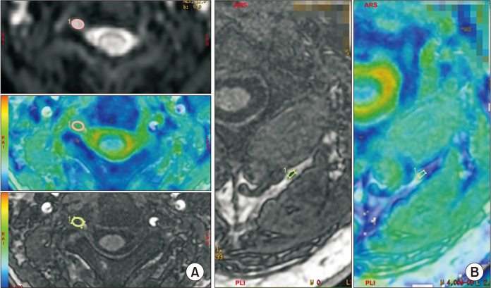
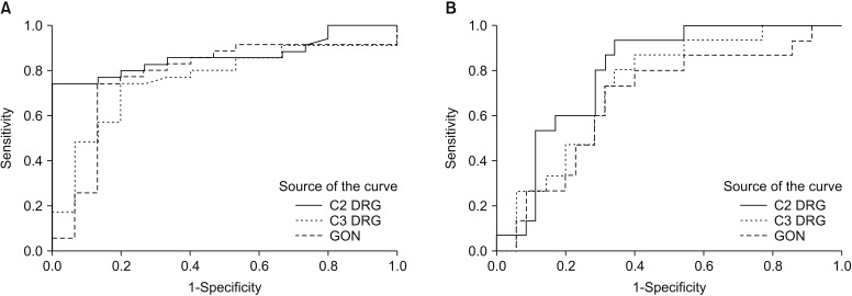
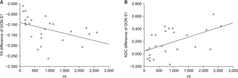
Methods: The fractional anisotropy (FA) and apparent diffusion coefficient (ADC) values in bilateral GONs and cervical DRG (C2 and C3) were measured. Grading values for headache severity was calculated using a receiver operating characteristics curve. The correlation was analyzed with Pearson's coefficient.

Results: The FA values of the symptomatic side of GON and cervical DRG (C2 and C3) were significantly lower than that of the asymptomatic side (all the P < 0.001), while the ADC values were significantly higher (P = 0.003, P < 0.001, and P = 0.003, respectively). The FA value of 0.205 in C2 DRG was considered the grading parameter for headache severity with sensitivity of 0.743 and specificity of 0.999 (P < 0.001). A negative correlation and a positive correlation between the FA and ADC value of the GON and headache index (HI; r = -0.420, P = 0.037 and r = 0.531, P = 0.006, respectively) was found.

Conclusions: DTI parameters in the symptomatic side of the C2 and C3 DRG and GON were significantly changed. The FA value of the C2 DRG can grade headache severity. DTI parameters of the GON significantly correlated with HI.

Keywords: Cervical Vertebrae; Chronic Pain; Diffusion Tensor Imaging; Ganglia; Headache; Magnetic Resonance Imaging; ROC Curve; Sensitivity and Specificity.; Spinal.


Conflict of interest statement

CONFLICT OF INTEREST

No potential conflict of interest relevant to this article was reported.

Figures

Fig. 1
Fig. 1
Multi-slice reconstruction on T2-weighted imaging CUBE showed the greater occipital nerve (GON) and muscles around. We firstly found the oblique capitis inferior muscle (white triangle) and its proximal inferior border (arrow). Then we recognized the proximal GON (arrowhead) as the target nerve for subsequent post-processing step.
Fig. 2
Fig. 2
Axial image showed the superimposed morphological T1-weighted imaging of right C3 dorsal root ganglia (DRG) (A) and left greater occipital nerve (GON) (B) with region of interest placement. The DRG was identified by directly visualization on original image (b = 0) and the target GON was identified with the help of aforementioned multi-slice reconstruction.
Fig. 3
Fig. 3
Fibre tractography showed the right greater occipital nerve (arrowheads) was thicker and disordered than the left (arrows) in a 48-year-old male with right cervicogenic headache for 1 month.
Fig. 4
Fig. 4
A tractography of a 24-year-old male healthy volunteer showed the fibre tract of bilateral side greater occipital nerves (GONs). The fibre tract of normal GON showed continuous, smooth and clearly visualization of all the segments (arrows).
Fig. 5
Fig. 5
A diagram about the enrollment of the patients. CGH: cervicogenic headache, CHISG: Cervicogenic Headache International Study Group, MRI: magnetic resonance imaging, GON: greater occipital nerve.
Fig. 6
Fig. 6
Graphs showed receiver operating characteristics (ROC) curves analysis of (A) fractional anisotropy and (B) apparent diffusion coefficient values, respectively. DRG: dorsal root ganglia, GON: greater occipital nerve.
Fig. 7
Fig. 7
Graphs showed the correlation between diffusion tensor imaging parameters of the nerves and HI. The fractional anisotropy (FA) value in proximal greater occipital nerve (GON) was negatively correlated with headache index (HI) (A), r = –0.420, P = 0.037. The apparent diffusion coefficient (ADC) value was positively correlated with HI (B), r = 0.531, P = 0.006.

References

    1. Yao X, Lin X. [Warming-needle moxibustion for cervical headache: a randomized controlled trial] Zhongguo Zhen Jiu. 2016;36:463–6. Chinese. - PubMed
    1. Wang E, Wang D. Treatment of cervicogenic headache with cervical epidural steroid injection. Curr Pain Headache Rep. 2014;18:442. doi: 10.1007/s11916-014-0442-3. - DOI - PMC - PubMed
    1. Pingree MJ, Sole JS, O’Brien TG, Eldrige JS, Moeschler SM. Clinical efficacy of an ultrasound-guided greater occipital nerve block at the level of C2. Reg Anesth Pain Med. 2017;42:99–104. doi: 10.1097/AAP.0000000000000513. - DOI - PubMed
    1. Zipfel J, Kastler A, Tatu L, Behr J, Kechidi R, Kastler B. Ultrasound-guided intermediate site greater occipital nerve infiltration: a technical feasibility study. Pain Physician. 2016;19:E1027–34. - PubMed
    1. Sapunar D, Kostic S, Banozic A, Puljak L. Dorsal root ganglion - a potential new therapeutic target for neuropathic pain. J Pain Res. 2012;5:31–8. doi: 10.2147/JPR.S26603. - DOI - PMC - PubMed

LinkOut - more resources