. 2020 Jun 30;11(6):490.
doi: 10.1038/s41419-020-2697-4.

Hfm1 participates in Golgi-associated spindle assembly and division in mouse oocyte meiosis

Affiliations

Hfm1 participates in Golgi-associated spindle assembly and division in mouse oocyte meiosis

Huiyuan Wang et al. Cell Death Dis. .

Abstract

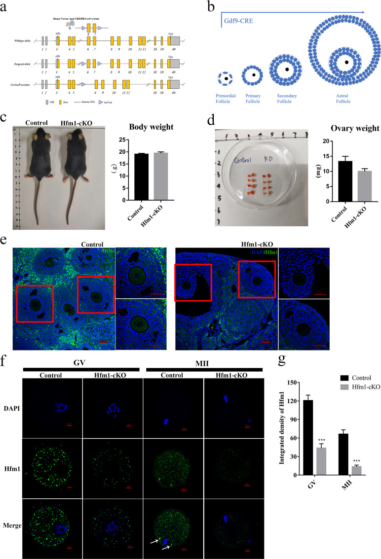
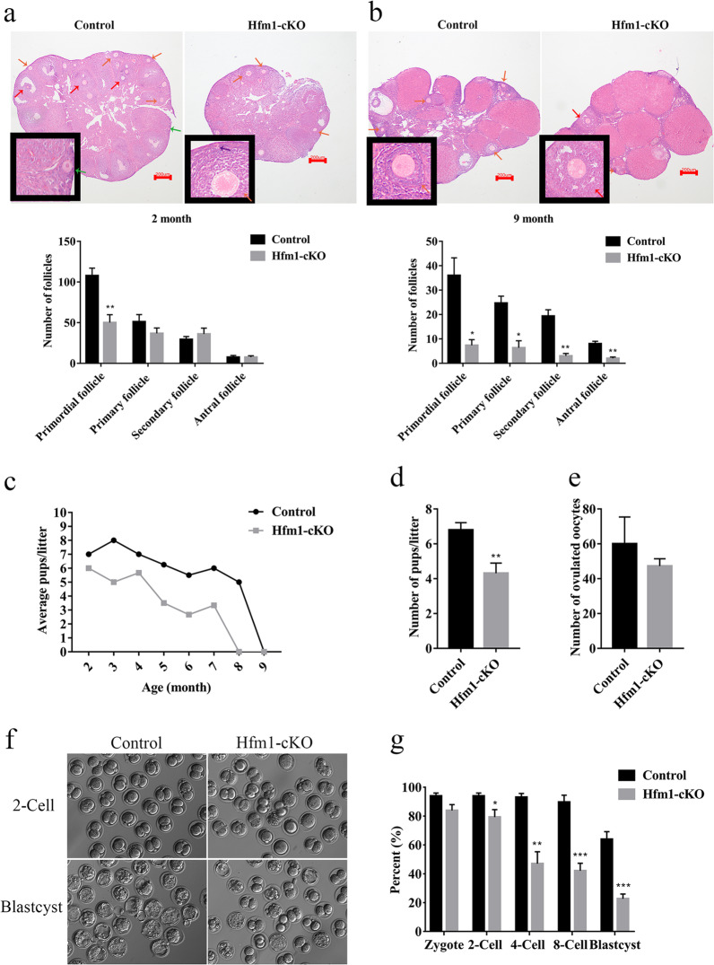
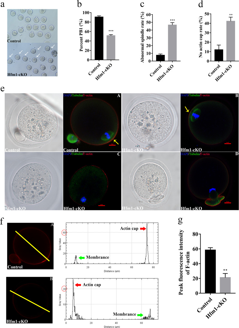
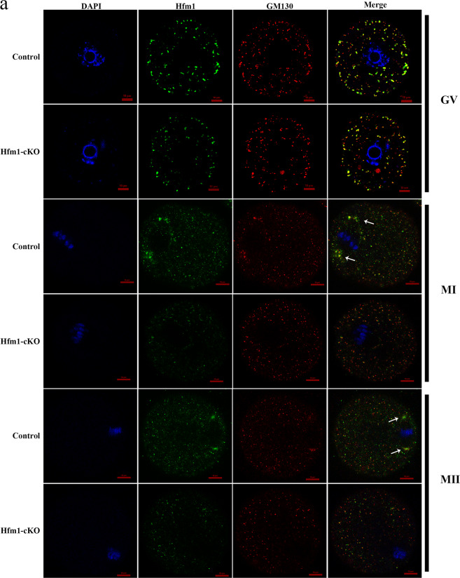
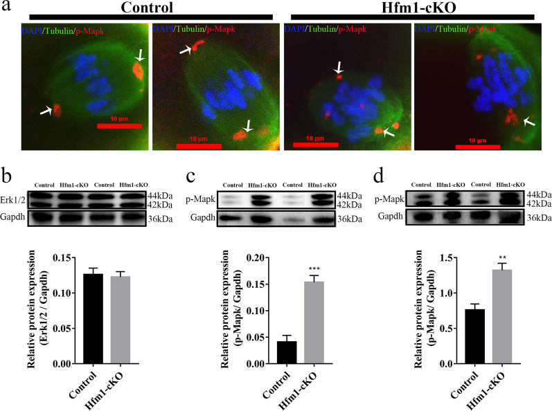
HFM1 (helicase for meiosis 1) is widely recognized as an ATP-dependent DNA helicase and is expressed mainly in germ-line cells. HFM1 is a candidate gene of premature ovarian failure (POF), hence it is also known as POF9. However, the roles of HFM1 in mammalian oocytes remain uncertain. To investigate the functions of HFM1, we established a conditional knockout (cKO) mouse model. Specific knockout of Hfm1 in mouse oocytes from the primordial follicle stage resulted in depletion of ovarian follicular reserve and subfertility of mice. In particular, abnormal spindle, misaligned chromosomes, loss of cortical actin cap, and failing polar body extrusion were readily observed in Hfm1-cKO oocytes. Further studies indicated that in addition to its cytoplasmic distribution, Hfm1 accumulated at the spindle poles, colocalized with the Golgi marker protein, GM130. Generally, GM130 signals overlapped with p-Mapk at the two spindle poles to regulate meiotic spindle assembly and asymmetric division. In this research, centrosome associated proteins, such as GM130 and p-Mapk, detached from the spindle poles in Hfm1-cKO oocytes. In conclusion, our data suggest that Hfm1 participates in Golgi-associated spindle assembly and division in mouse oocyte meiosis. These findings provide clues for pathogenesis of POF.


Conflict of interest statement

The authors declare that they have no competing interests.

Figures

Fig. 1
Fig. 1. Oocyte-specific Knockout of Hfm1 in mice.
a Engineered a conditional floxed allele for Hfm1 and a Cre-mediated recombination to delete exons 6 and 7. b Schematic illustration of Gdf9-Cre-mediated Hfm1 knockout in oocytes from primordial follicle stage c Photograph of whole bodies and measured the body weight of Control and Hfm1-cKO mice (2 months old) (n = 4). d Photograph of a pair of ovaries and measured the ovaries weight of Control and Hfm1-cKO mice (2-month-old) (n = 4). e Immunofluorescence staining of Hfm1 in ovaries of Control and Hfm1-cKO mice (2-month-old). f Immunofluorescence showed expression levels and subcellular localization of Hfm1 (green) in GV and MII oocytes of Control and Hfm1-cKO mice (2-month-old). DAPI are co-stained to visualize DNA (blue). White arrows point to spindle pole in oocytes. GV, Germinal Vesicle; MII, metaphase of meiosis II. g Quantification of the Hfm1 staining (n = 10). ***P < 0.01 by two-tailed Student’s t tests. Data represent the mean ± SEM.
Fig. 2
Fig. 2. Compromised fertility in Hfm1-cKO mice.
a H&E stained and follicle quantification in ovaries from adult Control and Hfm1-cKO mice (2-month-old) (n = 5). b H&E stained and follicle quantification in ovaries from older Control and Hfm1-cKO mice (9-month-old) (n = 4). The primordial (purple arrow), primary (green arrow), secondary (orange arrow) and antral follicles (red arrow) were annotated, and count data were presented per ovary. c Average pups numbers of each litter per month in Control and Hfm1-cKO mice (2-month-old) (n = 4). d The quantification analysis of pups per litter by the bar graph in a standard 7 months breeding trial. e Number of oocytes ovulated by prepubertal Control and Hfm1-cKO females (3-week-old) (n = 4). f Embryo developmental potential of oocytes before implantation from Control and Hfm1-cKO females (3-week-old) assessed by IVF. g The rate of zygote, 2-Cell, 4-Cell, 8-Cell, and blastocyst formation normalized to the number of MII oocytes ovulated by Control and Hfm1-cKO females (3-week-old) during IVF (n = 4). *P < 0.05. **P < 0.01. ***P < 0.001. Significance was determined by two-tailed Student’s t tests. Data represent the mean ± SEM.
Fig. 3
Fig. 3. Abnormal spindle in Hfm1 knockout oocytes.
a The micrographs of oocytes from Control and Hfm1-cKO females (3-week-old) after IVM. White arrows point to no first polar body (PB1) extrusion oocytes. b The PB1 extrusion rate of oocytes from Control and Hfm1-cKO females (3-week-old) by IVM (n = 4). c Percentage of in vivo matured MII oocytes with abnormal spindle from Control and Hfm1-cKO females (3-week-old) (n = 3). d Percentage of in vivo matured MII oocytes with no actin cap from Control and Hfm1-cKO females (3-week-old) (n = 3). e Representative spindles from MII oocytes ovulated by Control and Hfm1-cKO females (3-week-old) in vivo. (A) Normal MII stage. (B) abnormal MII with a spindle and a decreased actin cap fluorescence intensity. (C) Abnormal MII with no spindle and no actin cap. (D) Abnormal MII with incomplete division spindle and no actin cap. Microtubules, Chromosomes, and F-actin are stained green, blue, and red, respectively. Yellow arrows pointed to actin cap. f Profiles of phalloidin fluorescence intensity along the yellow line in oocytes (A) and (B). The yellow line perpendicular to the length axis of the spindle g Quantitative analysis of peak actin immunofluorescence intensities in profiles of Control and Hfm1-cKO oocytes with actin cap (n = 10). *P < 0.05. **P < 0.01. ***P < 0.001. Significance was determined by two-tailed Student’s t tests. Data represent the mean ± SEM.
Fig. 4
Fig. 4. Hfm1 functions in Golgi-associated spindle assembly and division.
a Double immunofluorescent staining of Hfm1 (green) and GM130 (red) in GV, MI and MII stage. DAPI are co-stained to visualize DNA (blue). White arrows point to spindle pole in oocytes. GV germinal vesicle, MI metaphase of meiosis I, MII metaphase of meiosis II.
Fig. 5
Fig. 5. Disrupted MAPK pathway in Hfm1 knockout oocytes.
a Denuded oocytes cultured for 8 h (MI) and stained with p-Mapk (red), β-Tubulin (green), and DAPI (blue). White arrows point to p-Mapk signals in oocytes. b Western blot analysis of Erk1/2 expression in Control and Hfm1-cKO ovaries (n = 5). c Western blot analysis of p-Mapk expression in Control and Hfm1-cKO ovaries (n = 5). d Western blot analysis of p-Mapk in Control and Hfm1-cKO oocytes at MI stage (n = 4). Protein lysates from 100 oocytes were loaded in each lane. **P < 0.01. ***P < 0.001. Significance was determined by two-tailed Student’s t tests. Data represent the mean ± SEM.

Similar articles

Cited by

References

    1. Tanaka K, Miyamoto N, Shouguchi-Miyata J, Ikeda JE. HFM1, the human homologue of yeast Mer3, encodes a putative DNA helicase expressed specifically in germ-line cells. DNA Seq. 2006;17:242–246. doi: 10.1080/10425170600805433. - DOI - PubMed
    1. European Society for Human, R. et al. ESHRE Guideline: management of women with premature ovarian insufficiency. Hum. Reprod. 2016;31:926–937. doi: 10.1093/humrep/dew027. - DOI - PubMed
    1. Tucker EJ, Grover SR, Bachelot A, Touraine P, Sinclair AH. Premature ovarian insufficiency: new perspectives on genetic cause and phenotypic spectrum. Endocr. Rev. 2016;37:609–635. doi: 10.1210/er.2016-1047. - DOI - PubMed
    1. Wang J, Zhang W, Jiang H, Wu BL, Primary Ovarian Insufficiency, C. Mutations in HFM1 in recessive primary ovarian insufficiency. N. Engl. J. Med. 2014;370:972–974. doi: 10.1056/NEJMc1310150. - DOI - PubMed
    1. Pu D, et al. Association analysis between HFM1 variation and primary ovarian insufficiency in Chinese women. Clin. Genet. 2016;89:597–602. doi: 10.1111/cge.12718. - DOI - PubMed

Publication types