

Tumor Cell Representation by an Improvised Technique of Fine-Needle Aspiration Specimen Acquisition and Cell Block Preparation: Our Experience in Lung Cancer Cases in a Peripheral Center of Eastern India
- PMID: 32606496
- PMCID: PMC7315915
- DOI: 10.4103/JOC.JOC_138_18
Tumor Cell Representation by an Improvised Technique of Fine-Needle Aspiration Specimen Acquisition and Cell Block Preparation: Our Experience in Lung Cancer Cases in a Peripheral Center of Eastern India
Abstract
Background: Being a minimally invasive diagnostic technique, Fine-Needle Aspiration Cytology (FNAC) has become the first-line test and corresponding aspirated material has become the target specimen for diagnosis and ancillary tests in lung carcinoma. Although the role of Cell Blocks (CBs) in diagnosis and in ancillary testing is well recognized in literature, limited attention has been paid to specimen procurement and triage in the preparation of CBs. In the present scenario, CBs are not consistently optimal because of its low cellularity.
Aims: This study is aimed to describe an improvised technique of specimen acquisition and cell block preparation in CT-guided FNACs of lung carcinoma cases in a resource-constrained center and to assess its efficacy for optimal representation of cellularity, morphology, and architecture.
Materials and methods: Total 85 lung carcinoma cases undergoing CT-guided FNAC in our center from February 2017 to January 2018 were included in this study. 4 to 5 direct smears and subsequent CBs were made from material obtained by single pass. Cellularity of smears and corresponding cell blocks were assessed and categorized according to a scoring system (score 1 to 3 for number of cells <50, 50-100, >100, respectively). Preserved architecture and morphology were also assessed in smears and CBs.
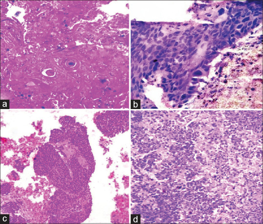
Results: The evaluated samples showed a cellularity score 3 in 65.4%CBs and score 2 in 24.7% CBs. Overall, 90.1% cell blocks had acceptable cellularity. Cell morphology was preserved in all CBs of acceptable cellularity, except for two adenocarcinoma, one squamous cell carcinoma, and one small cell carcinoma blocks. Cellular architecture was also preserved in all CBs of acceptable cellularity.
Conclusions: This simple improvised technique of CB preparation optimized its cellularity, morphology, and architectural preservation, even after adequate cellular FNA smears.
Keywords: Architecture; cell block; cellularity; fine-needle aspiration cytology; lung cancer; morphology.
Copyright: © 2020 Journal of Cytology.
Conflict of interest statement
There are no conflicts of interest.
Figures

Similar articles
-
Appreciation of Pattern in Diagnosis of Lung Adenocarcinoma from Cytology Specimen: Our Experience with Fine Needle Aspiration Cytology and Cell Block in a Resource Constraint Setup.J Cytol. 2020 Jul-Sep;37(3):141-146. doi: 10.4103/JOC.JOC_148_19. Epub 2020 Jul 10. J Cytol. 2020. PMID: 33088033 Free PMC article.
-
The utility of cellient cell blocks in low-cellularity thyroid fine needle aspiration biopsies.Diagn Cytopathol. 2016 Sep;44(9):737-41. doi: 10.1002/dc.23522. Epub 2016 Jun 24. Diagn Cytopathol. 2016. PMID: 27338858
-
Utility of cell blocks in the diagnosis of thyroid aspirates.Diagn Cytopathol. 2006 Feb;34(2):89-92. doi: 10.1002/dc.20385. Diagn Cytopathol. 2006. PMID: 16514670
-
Fine Needle Aspiration Cytology's Role in the Diagnosis of Ovarian Tumor.J Midlife Health. 2023 Jul-Sep;14(3):159-164. doi: 10.4103/jmh.jmh_82_23. Epub 2023 Dec 30. J Midlife Health. 2023. PMID: 38312758 Free PMC article. Review.
-
Cell blocks in cytopathology: An update.Cytopathology. 2018 Dec;29(6):505-524. doi: 10.1111/cyt.12627. Epub 2018 Sep 27. Cytopathology. 2018. PMID: 30153355 Review.
Cited by
-
Appreciation of Pattern in Diagnosis of Lung Adenocarcinoma from Cytology Specimen: Our Experience with Fine Needle Aspiration Cytology and Cell Block in a Resource Constraint Setup.J Cytol. 2020 Jul-Sep;37(3):141-146. doi: 10.4103/JOC.JOC_148_19. Epub 2020 Jul 10. J Cytol. 2020. PMID: 33088033 Free PMC article.
References
-
- Siegel R, Naishadham D, Jemal A. Cancer statistics, 2013. CA Cancer J Clin. 2013;63:11–30. - PubMed
-
- Warth A, Muley T, Meister M, Stenzinger A, Thomas M, Schirmacher P, et al. The novel histologic International Association for the Study of Lung Cancer/American Thoracic Society/European Respiratory Society classification system of lung adenocarcinoma is a stage-independent predictor of survival. J Clin Oncol. 2012;30:1438–46. - PubMed
-
- Warth A, Muley T, Herpel E, Meister M, Herth FJ, Schirmacher P, et al. Large-scale comparative analyses of immunomarkers for diagnostic subtyping of non-small-cell lung cancer biopsies. Histopathology. 2012;61:1017–25. - PubMed
-
- Rivera MP, Mehta AC, Wahidi MM. Establishing the diagnosis of lung cancer: Diagnosis and management of lung cancer, 3rd ed: American College of Chest Physicians evidence-based clinical practice guidelines. Chest. 2013;143(Suppl 5):e142S–165S. - PubMed
-
- Kossakowski C, Morresi-Hauf A, Schnabel P, Eberhardt R, Herth F, Warth A. Preparation of cell blocks for lung cancer diagnosis and prediction: Protocol and experience of a high-volume center. Respiration. 2014;87:432–8. - PubMed
LinkOut - more resources
Full Text Sources