

Fine-Needle Cytological Characteristics of Carcinoma Breast with Medullary or Medullary-like Features Masquerading as Dendritic Reticulum Cell Sarcoma: An Attempt to Explore the Reasons for Erroneous Cytologic Interpretation
- PMID: 32606498
- PMCID: PMC7315914
- DOI: 10.4103/JOC.JOC_15_20
Fine-Needle Cytological Characteristics of Carcinoma Breast with Medullary or Medullary-like Features Masquerading as Dendritic Reticulum Cell Sarcoma: An Attempt to Explore the Reasons for Erroneous Cytologic Interpretation
Abstract
Background: Infiltration of tumors by dendritic reticulum cells (DRC) reflects the host immune defense mechanism. We observed three breast carcinomas cases with dense tumor-infiltrating DRC and lymphocytes in fine-needle aspiration (FNA) smears, leading to cytodiagnosis or differential diagnosis of dendritic reticulum cell sarcoma (DRCS). An attempt was made to find out the reason behind such an erroneous interpretation.
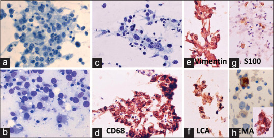
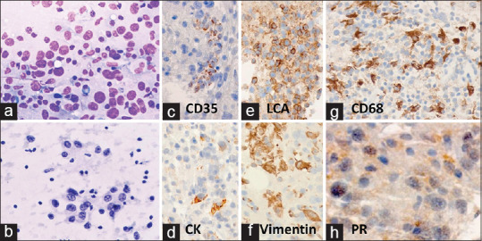
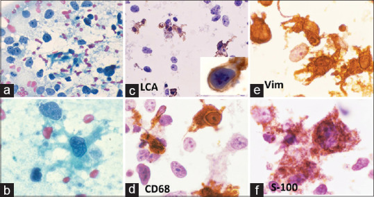
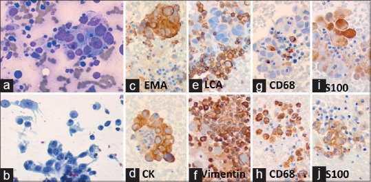
Materials and methods: Between 2009 and 2014, two cases were diagnosed as DRCS of the female breast by FNA cytology and in one case possibility of DRCS was considered along with medullary breast carcinoma (MBC). We compare and contrast the cytomorphological features of these three cases with those of nine cytologically diagnosed MBC.
Results: Cases diagnosed as DRCS or MBC showed singly dispersed tumor cells, nuclear pleomorphism, bare nuclei, prominent nucleoli, and presence of lymphocytes. There was no significant difference between the two groups for discohesive clusters, syncytial clusters, plasma cells, neutrophils, foamy histiocytes, and necrosis. However, there was significant difference for presence of cohesive clusters (0% DRCS and 100% MBC, P = 0.00485), severe degree (+++) of pleomorphism (100% DRCS vs. 11.1% MBC, P = 0.01818), +++ DRC (P = 0.04697), and DRC with ++ to +++ enlarged nuclei (P = 0.03333), and pleomorphic nuclei (P = 0.00833). Two of the three cytologically diagnosed DRCS cases proved to be MBC or MBC-like and one as invasive ductal carcinoma. Six of nine cytologically diagnosed MBC cases with histology proved to be invasive breast carcinomas.
Conclusion: Criteria for cytodiagnosis MBC need a fresh look. Cases with numerous dendritic cells possibly represent MBC.
Keywords: Breast; Medullary-like carcinoma; dendritic reticulum cell sarcoma; fine-needle aspiration cytology; medullary carcinoma.
Copyright: © 2020 Journal of Cytology.
Conflict of interest statement
There are no conflicts of interest.
Figures
Similar articles
-
Fine-needle aspiration cytologic features of four special types of breast cancers: mucinous, medullary, apocrine, and papillary.Diagn Cytopathol. 2007 Jul;35(7):408-16. doi: 10.1002/dc.20661. Diagn Cytopathol. 2007. PMID: 17580344
-
Cytomorphological Study of Medullary Carcinoma of Breast in Comparison to Infiltrating Ductal Carcinoma.J Cytol. 2018 Oct-Dec;35(4):195-198. doi: 10.4103/JOC.JOC_160_17. J Cytol. 2018. PMID: 30498288 Free PMC article.
-
Cytodiagnosis of papillary carcinoma of the breast: Report of a case with histological correlation.J Cytol. 2014 Apr;31(2):119-21. doi: 10.4103/0970-9371.138694. J Cytol. 2014. PMID: 25210247 Free PMC article.
-
Fine-needle aspiration cytology of medullary breast carcinoma: report of two cases and review of the literature with emphasis on differential diagnosis.Diagn Cytopathol. 2007 Jun;35(6):313-8. doi: 10.1002/dc.20639. Diagn Cytopathol. 2007. PMID: 17497662 Review.
-
Fine-needle aspiration of epithelioid sarcoma: cytology findings in nine cases.Cancer. 2001 Aug 25;93(4):246-51. doi: 10.1002/cncr.9037. Cancer. 2001. PMID: 11507697 Review.
References
-
- Kapucuoglu N, Percinel S, Ventura T, Lang R, Al-Daraji W, Eusebi V. Dendritic cell sarcoma/tumors of the breast: Report of two cases. Virchows Arch. 2009;454:333–9. - PubMed
-
- Andriko JW, Kaldjian EP, Tsokos M, Abbondanzo SL, Jaffe ES. Reticulum cell neoplasms of lymph nodes: A clinicopathologic study of 11 cases with recognition of a new subtype derived from fibroblastic reticular cells. Am J Surg Pathol. 1998;22:1048–58. - PubMed
-
- Wright-Browne V, McClain KL, Talpaz M, Ordonez N, Estrov Z. Physiology and pathophysiology of dendritic cells. Hum Pathol. 1997;28:563–79. - PubMed
-
- Lespagnard L, Gancberg D, Rougas G, Leclercq G, de Saint-Aubain Somerhausen N, Di Leo A, et al. Tumor-infiltrating dendritic cells in adenocarcinomas of the breast: A study of 143 neoplasms with a correlation to usual prognostic factors and to clinical outcome. Int J Cancer. 1999;84:309–14. - PubMed
LinkOut - more resources
Full Text Sources