. 2020 Jun 12:13:37-45.
doi: 10.2147/NSA.S254770. eCollection 2020.

Cordycepin Nanoencapsulated in Poly(Lactic-Co-Glycolic Acid) Exhibits Better Cytotoxicity and Lower Hemotoxicity Than Free Drug

Affiliations

Cordycepin Nanoencapsulated in Poly(Lactic-Co-Glycolic Acid) Exhibits Better Cytotoxicity and Lower Hemotoxicity Than Free Drug

Gregory Marslin et al. Nanotechnol Sci Appl. .

Abstract

Purpose: Cordycepin, a natural product isolated from the fungus Cordyceps militaris, is a potential candidate for breast cancer therapy. However, due to its structural similarity with adenosine, cordycepin is rapidly metabolized into an inactive form in the body, hindering its development as a therapeutic agent. In the present study, we have prepared cordycepin as nanoparticles in poly(lactic-co-glycolic acid) (PLGA) and compared their cellular uptake, cytotoxicity and hemolytic potential with free cordycepin.

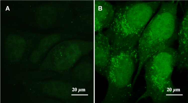
Materials and methods: Cordycepin-loaded PLGA nanoparticles (CPNPs) were prepared by the double-emulsion solvent evaporation method. Physico-chemical characterization of the nanoparticles was done by zetasizer, transmission electron microscopy (TEM) and reverse-phase high-pressure liquid chromatography (RP-HPLC) analyses. Cellular uptake and cytotoxicity of CPNPs and free drug were tested in human breast cancer cells (MCF7). Hemolytic potential of both of these forms was evaluated in rat red blood cells (RBCs).

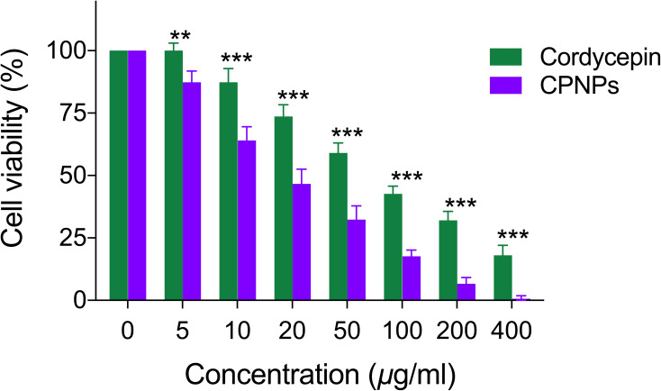
Results: Physico-chemical characterization revealed that CPNPs were spherical in shape, possessed a size range of 179-246 nm, and released the encapsulated drug sustainably over a period of 10 days. CPNPs exhibited a high level of cellular uptake and cytotoxicity than the free drug in MCF-7 cells. While CPNPs were not toxic to rat RBCs even at high concentrations, free cordycepin induced hemolysis of these cells at relatively low concentration.

Conclusion: Our results reveal that delivery as CPNPs could enhance the clinical efficacy of cordycepin substantially.

Keywords: PLGA; breast cancer cells; cellular uptake; cordycepin; cytotoxicity; hemolysis; nanoparticles.


Conflict of interest statement

The authors report no conflicts of interest in this work.

Figures

Figure 1
Figure 1
Structure of cordycepin (A) and adenosine (B) obtained from the Pubchem database showing similarity.
Figure 2
Figure 2
(A) Transmission electron micrograph showing cordycepin-loaded PLGA nanoparticles (CPNPs) of C3 formulation and (B) Histogram showing the size distribution of CPNPs in the formulation.
Figure 3
Figure 3
HPLC chromatogram of standard cordycepin (A), UV spectrum of standard cordycepin (B), HPLC chromatogram of cordycepin extracted from CPNPs (C) and, UV spectrum of cordycepin extracted from CPNPs (D).
Figure 4
Figure 4
In vitro release profile of cordycepin from CPNPs in PBS at pH 7.4 (mean ± SD, n = 3). Statistical analysis of data was performed using two-way ANOVA, followed by Bonferroni post hoc test. C2 vs C3: *p<0.05, ***p<0.001; C1 vs C2: $p<0.05; C2 vs C4: #p<0.05.
Figure 5
Figure 5
Comparison of cytotoxic potential of free cordycepin and CPNP in MCF-7 cells (mean ± SD, n = 3). Statistical analysis of data was performed using two-way ANOVA, followed by Bonferroni post hoc test. Cordycepin vs CPNPs (**p<0.01, ***p<0.001).
Figure 6
Figure 6
Uptake of curcumin (A) and curcumin loaded PLGA nanoparticles (B) by MCF-7 cells after 4 h of incubation as revealed by green fluorescence under the confocal microscope.
Figure 7
Figure 7
Effect of free cordycepin and CPNPs on the hemolysis of in rat RBCs. (A) distilled water; (B) normal saline; (C) 50 µg/mL free cordycepin; (D) 100 µg/mL free cordycepin; (E) 100 µg/mL CPNPs.

References

    1. Shi P, Huang Z, Tan X, Chen G. Proteomic detection of changes in protein expression induced by cordycepin in human hepatocellular carcinoma BEL-7402 cells. Methods Find Exp Clin Pharmacol. 2008;30:347–353. doi:10.1358/mf.2008.30.5.1186085 - DOI - PubMed
    1. Li G, Nakagome I, Hirono S, Itoh T, Fujiwara R. Inhibition of adenosine deaminase (ADA) mediated metabolism of cordycepin by natural substances. Pharmacol Res Perspect. 2015;3:e00121. doi:10.1002/prp2.121 - DOI - PMC - PubMed
    1. Cao T, Xu R, Xu Y, Liu Y, Qi D, Wan Q. The protective effect of Cordycepin on diabetic nephropathy through autophagy induction in vivo and in vitro. Int Urol Nephrol. 2019;51:1883–1892. doi:10.1007/s11255-019-02241-y - DOI - PubMed
    1. Ashraf S, Radhi M, Gowler P, et al. The polyadenylation inhibitor cordycepin reduces pain, inflammation and joint pathology in rodent models of osteoarthritis. Sci Rep. 2019;9:4696. doi:10.1038/s41598-019-41140-1 - DOI - PMC - PubMed
    1. Cho HJ, Cho JY, Rhee MH, Kim HS, Lee HS, Park HJ. Inhibitory effects of cordycepin (3ʹ-deoxyadenosine), a component of Cordyceps militaris, on human platelet aggregation induced by thapsigargin. J Microbiol Biotechnol. 2007;17:1134–1138. - PubMed

LinkOut - more resources