. 2020 Jun 11:15:4079-4090.
doi: 10.2147/IJN.S250960. eCollection 2020.

Formulation and Characterization of Sertaconazole Nitrate Mucoadhesive Liposomes for Vaginal Candidiasis

Affiliations

Formulation and Characterization of Sertaconazole Nitrate Mucoadhesive Liposomes for Vaginal Candidiasis

Menna M Abdellatif et al. Int J Nanomedicine. .

Abstract

Purpose: The aim of this study is to develop efficient localized therapy of sertaconazole nitrate for the treatment of vaginal candidiasis.

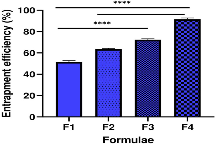
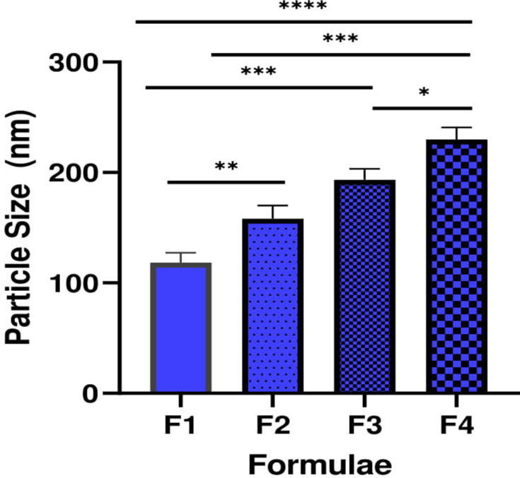
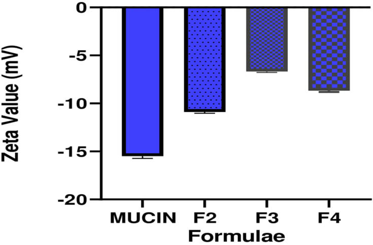
Methods: Sertaconazole nitrate-loaded cationic liposomes were prepared by thin-film hydration method and coated with different concentrations of pectin (0.05%, 0.1% and 0.2%) to develop mucoadhesive liposomes. The formulated mucoadhesive vesicles were characterized in terms of morphology, entrapment efficiency, particle size, zeta value, mucoadhesive properties and drug release. The selected formula was incorporated into a gel base and further characterized by an ex vivo permeation study in comparison with conventional sertaconazole gel. Also, the in vivo study was performed to assess the efficacy of sertaconazole mucoadhesive liposomal gel in treating rats with vaginal candidiasis.

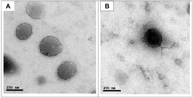
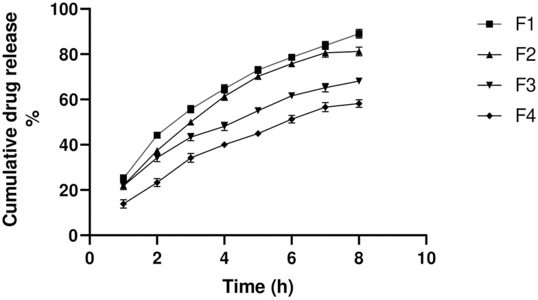
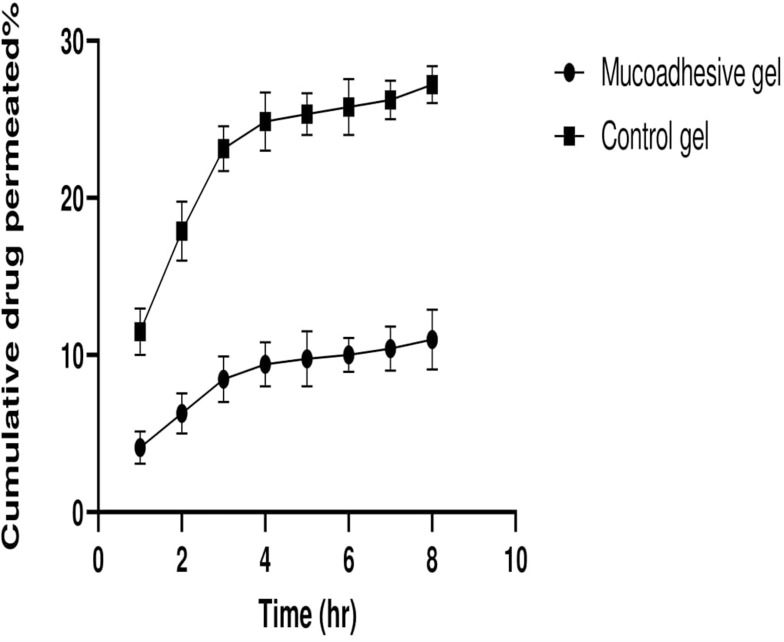
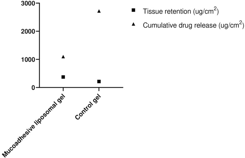
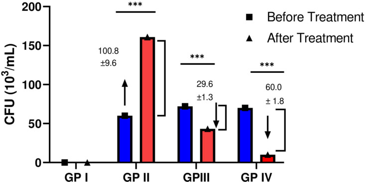
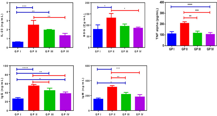
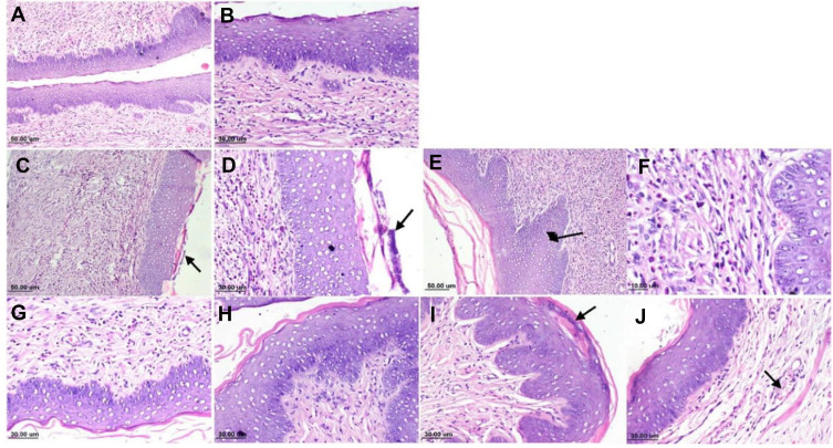
Results: The mucoadhesive liposomes were spherical. Coating liposomes with pectin results in increased entrapment efficiency and particle size compared with uncoated vesicles. On the contrary, zeta values were reduced upon coating liposomes with pectin indicating efficient coating of liposomes with pectin. Mucoadhesive liposomes showed a more prolonged and sustained drug release compared with uncoated liposomes. Ex vivo study results showed that mucoadhesive liposomal gel increased sertaconazole tissue retention and reduced drug tissue penetration. In the invivo study, the mucoadhesive liposomal gel showed a significant reduction in the microbial count with a subsequent reduction in inflammatory responses with the lowest histopathological change compared with conventional gel.

Conclusion: The study confirmed the potentiality of employing mucoadhesive liposomes as a successful carrier for the vaginal delivery of antifungal drugs.

Keywords: mucoadhesive liposomes; sertaconazole nitrate; vaginal candidiasis.


Conflict of interest statement

The authors report no conflicts of interest in this work.

Figures

Figure 1
Figure 1
TEM images of the sertaconazole-loaded vesicles displaying (A) liposomes, (B) mucoadhesive liposomes.
Figure 2
Figure 2
Entrapment efficiency of different formulae (n=3). ****P < 0.0001.
Figure 3
Figure 3
Particle size diameter of different formulae (n=3). *P < 0.05, **P < 0.01, ***P < 0.001, and ****P < 0.0001.
Figure 4
Figure 4
Zeta potential shift of mucin upon mixing with different mucoadhesive liposomes.
Figure 5
Figure 5
In vitro drug release profile.
Figure 6
Figure 6
Ex vivo permeation drug profile.
Figure 7
Figure 7
Cumulative drug release and tissue retention of mucoadhesive liposomal gel and control gel.
Figure 8
Figure 8
Mean colony-forming unit (CFU) of each group before and after the treatment period (n=6). ***P < 0.001.
Figure 9
Figure 9
Assessment of serum Interleukin-23 (IL-23), beta-D-glucan (BDG), tumor necrosis factor-alpha (TNF-α), Immunoglobulin G (IgG) and Immunoglobulin M (IgM) (n=6). *P < 0.05, **P < 0.01, ***P < 0.001, and ****P < 0.0001.
Figure 10
Figure 10
Photomicrograph of rat vagina, H&E stain: (A) Negative control group, normal histology of rat vagina; (B) negative control group, higher magnification, showing stratified squamous epithelium with dense sub-epithelial connective tissue; (C) positive control group, showing heavy subepithelial inflammatory cells infiltration with necrotic debris over the mucosal surface (arrow); (D) positive control group, higher magnification, showing dissolution of keratin layer with presence of necrotic tissue debris; (E) positive control group, showing hyperplastic mucosa (arrow); (F) positive control group, showing sub-epithelial neutrophils and mononuclear infiltration; (G) mucoadhesive liposomal gel group, showing mild sub-epithelial inflammatory cells infiltration; (H) showing intact mucosa; (I) sertaconazole control gel, showing mucosa with presence of necrotic debris in the keratin layer and (J) showing mild sub-epithelial edema with dilated blood vessels (arrow).

References

    1. Albertini B, Passerini N, Di Sabatino M, Vitali B, Brigidi P, Rodriguez L. Polymer–lipid based mucoadhesive microspheres prepared by spray-congealing for the vaginal delivery of econazole nitrate. Eur J Pharm Sci. 2009;36(4–5):591–601. doi:10.1016/j.ejps.2008.12.009 - DOI - PubMed
    1. Hainer BL, Gibson MV. Vaginitis: diagnosis and treatment. Am Fam Physician. 2011;83(7):807–815. - PubMed
    1. Vanić Ž, Hurler J, Ferderber K, Golja Gašparović P, Škalko-Basnet N, Filipović-Grčić J. Novel vaginal drug delivery system: deformable propylene glycol liposomes-in-hydrogel. J Liposome Res. 2014;24(1):27–36. doi:10.3109/08982104.2013.826242 - DOI - PubMed
    1. Takeuchi H, Yamamoto H, Kawashima Y. Mucoadhesive nanoparticulate systems for peptide drug delivery. Adv Drug Deliv Rev. 2001;47(1):39–54. doi:10.1016/S0169-409X(00)00120-4 - DOI - PubMed
    1. Netsomboon K, Bernkop-Schnürch A. Mucoadhesive vs. mucopenetrating particulate drug delivery. Eur J Pharm Biopharm. 2016;98:76–89. doi:10.1016/j.ejpb.2015.11.003 - DOI - PubMed

MeSH terms