. 2020 Jun 19:15:4333-4350.
doi: 10.2147/IJN.S254342. eCollection 2020.

An AFM-Based Nanomechanical Study of Ovarian Tissues with Pathological Conditions

Affiliations

An AFM-Based Nanomechanical Study of Ovarian Tissues with Pathological Conditions

Arian Ansardamavandi et al. Int J Nanomedicine. .

Abstract

Background: Different diseases affect both mechanical and chemical features of the involved tissue, enhancing the symptoms.

Methods: In this study, using atomic force microscopy, we mechanically characterized human ovarian tissues with four distinct pathological conditions: mucinous, serous, and mature teratoma tumors, and non-tumorous endometriosis. Mechanical elasticity profiles were quantified and the resultant data were categorized using K-means clustering method, as well as fuzzy C-means, to evaluate elastic moduli of cellular and non-cellular parts of diseased tissues and compare them among four disease conditions. Samples were stained by hematoxylin-eosin staining to further study the content of different locations of tissues.

Results: Pathological state vastly influenced the mechanical properties of the ovarian tissues. Significant alterations among elastic moduli of both cellular and non-cellular parts were observed. Mature teratoma tumors commonly composed of multiple cell types and heterogeneous ECM structure showed the widest range of elasticity profile and the stiffest average elastic modulus of 14 kPa. Samples of serous tumors were the softest tissues with elastic modulus of only 400 Pa for the cellular part and 5 kPa for the ECM. Tissues of other two diseases were closer in mechanical properties as mucinous tumors were insignificantly stiffer than endometriosis in cellular part, 1300 Pa compared to 1000 Pa, with the ECM average elastic modulus of 8 kPa for both.

Conclusion: The higher incidence of carcinoma out of teratoma and serous tumors may be related to the intense alteration of mechanical features of the cellular and the ECM, serving as a potential risk factor which necessitates further investigation.

Keywords: atomic force microscopy; cellular and extra cellular matrix components; ovarian tumors; tissue elasticity.


Conflict of interest statement

The authors report no conflicts of interest in this work.

Figures
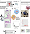
Figure 1
Figure 1
A representation of experimental approach. (A) Schematic representation of the study. (B) AFM measurement of the mature teratoma ovarian tissue sample, the tissue was tested while adhered to a graded plate to identify the approximate location of AFM measurement with a specific dimension of (0.3 × 1 × 1.5 cm) in DMEM solution. (C) The obtained data of AFM measurements. AFM tip pushes down the tissue containing cells and ECM documenting the indentation depth (μm) versus force (nN) generated by pushing down the section. This result was used for obtaining elasticity according to the fundamental equations. (D) The image represents the sample which was prepared for histopathological staining after AFM indentation (top), and the histopathological stained section of mature teratoma (bottom).
Figure 2
Figure 2
Represents Young’s modulus histogram of tissues of different ovarian diseases in logarithmic scale with a Gaussian distribution plot fitted on histograms. (A) Endometriosis, (B) mucinous cystadenoma, (C) serous cystadenoma, (D) mature teratoma, and (E) the average elastic moduli of ovarian tissues with four diseases. Measurements were carried out by atomic force microscopy in force spectroscopy mode at room temperature under physiological conditions. The significant differences in parameters of test groups were reported with the * sign (****p<0.0001).
Figure 3
Figure 3
Obtaining number of clusters by elbow methods. Figure represents average within the cluster distance of elasticity profiles of indented tissues four ovarian diseases versus the clustering number. (A) Elbow method for endometriosis. (B) Elbow method for mucinous cystadenoma. (C) Elbow method for serous cystadenoma. (D) Elbow method for mature teratoma. Results were further verified by silhouette method.
Figure 4
Figure 4
Represents the cellular elasticity profiles of ovarian tissues of four different diseases measured by atomic force microscopy. (A) Endometriosis, (B) mucinous cystadenoma, (C) serous cystadenoma, (D) mature teratoma. K-means clustering was applied to categorize elasticity data. K-means clustering center 1 (kmeanC1) represents the clustering center of cellular region which significantly depends on the disease condition.
Figure 5
Figure 5
Represents the average elastic moduli of cellular and ECM parts of quantified diseases. (A) Cellular and (B–D) extracellular matrix portions. Bonferroni’s multiple comparison test was performed to compare data statistically. The significant differences in parameters of test groups were reported with the *sign (*p<0.05, **p<0.01, and ****p<0.0001).
Figure 6
Figure 6
Histopathological examination of ovarian tissues. (A and B) Microscopic examination of hematoxylin and eosin (H&E) stained sections demonstrate endometriosis. (A) Microscopic low power examination (10×) reveals ovarian cyst lined by endometrial glands and decasualized stroma is evident. Hemosiderin laden macrophages and features of old hemorrhage are also seen. (B) High-power examination (40×) of the stromal cells shows abundant eosinophilic cytoplasm, distinct cell membrane, and round uniform nuclei consistent with progesterone hormonal effect on the underlying endometriosis. (C and D) Microscopic examination of hematoxylin and eosin (H&E) stained sections demonstrate mucinous cystadenoma. (C) Microscopic low power examination (10×) shows ovarian tissue with cystic structure, fibrotic stroma and (D) microscopic high-power examination (40×) shows a flat mucinous epithelial lining, without significant atypia compatible with mucinous cystadenoma.
Figure 7
Figure 7
Histopathological examination of ovarian tissues. (A and B) Microscopic examination of hematoxylin and eosin (H&E) stained sections demonstrate serous cystadenoma. (A) Microscopic low power examination (10×) shows ovarian tissue with fibrotic stroma and multiple cystic structures. (B) Microscopic high-power examination (40×) depicts that the tissue sections were lined with flat or cuboidal ciliated epithelial cells without significant atypia compatible with serous cystadenoma. (C and D) Microscopic examination of hematoxylin and eosin (H&E) stained sections demonstrate mature teratoma. (C) Microscopic low power examination (10×) reveals ovarian cyst with keratinizing squamous epithelial lining, normal skin appendices, and hair follicle consistent with mature teratoma. (D) High-power examination (40×) reveals a mature adipose tissue (at the bottom) and sebaceous gland (left of the figure) are seen.

References

    1. Kai F, Laklai H, Weaver VM. Force matters: biomechanical regulation of cell invasion and migration in disease. Trends Cell Biol. 2016;26(7):486–497. doi:10.1016/j.tcb.2016.03.007 - DOI - PMC - PubMed
    1. Alibert C, Goud B, Manneville JB. Are cancer cells really softer than normal cells? Biol Cell. 2017;109(5):167–189. doi:10.1111/boc.201600078 - DOI - PubMed
    1. Kraning-Rush CM, Califano JP, Reinhart-King CA. et al. Cellular traction stresses increase with increasing metastatic potential. PLoS One. 2012;7(2):e32572. doi:10.1371/journal.pone.0032572 - DOI - PMC - PubMed
    1. Ansardamavandi A, Tafazzoli-Shadpour M, Omidvar R, Jahanzad I. Quantification of effects of cancer on elastic properties of breast tissue by atomic force microscopy. J Mech Behav Biomed Mater. 2016;60:234–242. doi:10.1016/j.jmbbm.2015.12.028 - DOI - PubMed
    1. Cross SE, Jin Y-S, Rao J, Gimzewski JK. Nanomechanical analysis of cells from cancer patients. Nat Nanotechnol. 2007;2(12):780. doi:10.1038/nnano.2007.388 - DOI - PubMed