. 2020 Jun 23:15:4501-4521.
doi: 10.2147/IJN.S255392. eCollection 2020.

Anti-Oxidant and Anti-Endothelial Dysfunctional Properties of Nano-Selenium in vitro and in vivo of Hyperhomocysteinemic Rats

Affiliations

Anti-Oxidant and Anti-Endothelial Dysfunctional Properties of Nano-Selenium in vitro and in vivo of Hyperhomocysteinemic Rats

Zeqi Zheng et al. Int J Nanomedicine. .

Abstract

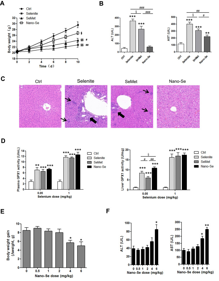
Purpose: Elevation of blood homocysteine (Hcy) level (hyperhomocysteinemia) is a risk factor for cardiovascular disorders and is closely associated with endothelial dysfunction. The present study aims to investigate the protective effect and underlying mechanism of nanoscale selenium (Nano-Se) in Hcy-mediated vascular endothelial cell dysfunction in vitro and in vivo.

Materials and methods: By incubating vascular endothelial cells with exogenous Hcy and generating hyperhomocysteinemic rat model, the effects of Nano-Se on hyperhomocysteinemia-mediated endothelial dysfunction and its essential mechanisms were investigated.

Results: Nano-Se inhibited Hcy-induced mitochondrial oxidative damage and apoptosis by preventing the downregulation of glutathione peroxidase enzyme 1 and 4 (GPX1, GPX4) in the vascular endothelial cells, thus effectively prevented the vascular damage in vitro and in vivo in the hyperhomocysteinemic rats. Nano-Se possessed similar protective effects but lower toxicity against Hcy in vascular endothelial cells when compared with other forms of Se.

Conclusion: The application of Nano-Se could serve as a novel promising strategy against Hcy-mediated vascular dysfunction with reduced risk of Se toxicity.

Keywords: GPXs; Nano-Se; ROS; endothelium dysfunction; glutathione peroxidase enzymes; homocysteine; nano-selenium; reactive oxygen species.


Conflict of interest statement

The authors report no conflicts of interest in this work.

Figures

Figure 1
Figure 1
Nano-Se protected HUVECs against Homocysteine-mediated apoptosis. (A) Transmission electron microscopy image of Nano-Se. (B) Dynamic light scattering measurement. (C) Dose-dependent effect of Nano-Se on HUVEC viability with Hcy. The cells were pre-treated with 100–700 nM Nano-Se for 8 hours before incubation with 1.5 mM Hcy for 24 hours. Cell viability was analyzed by LDH release assay. The histograms show the mean ± SEM of three separate experiments, each measured in triplicate (§§p<0.01 vs Ctrl; *p<0.05, **p<0.01 vs Hcy treatment; #p<0.05 and ##p<0.01 vs Hcy+100 nM Nano-Se treatment; &p<0.05 vs Hcy+300 nM Nano-Se treatment). (D) Caspase 3 activation in HUVECs treated with Hcy in the presence or absence of Nano-Se. The cells were pre-treated with 500 nM Nano-Se for 8 hours before incubation with 1.5 mM Hcy for 20 hours. Caspase 3 activation was detected by flow cytometry analysis. The numbers indicate the gating of a subpopulation of cells with positive caspase 3 activation. Representative histograms of three separate experiments are shown. The numbers indicate the gating of the subpopulation of cells with positive caspase 3 activation. (E) Determination of mitochondrial cytochrome c in HUVECs with 1.5 mM Hcy in the presence or absence of 500 nM Nano-Se for 20h, detected by flow cytometry analysis. The cells were pre-treated with 500 nM Nano-Se 8 hours before 1.5 mM Hcy was added. The overlays show the distribution of mitochondrial cytochrome c fluorescence intensity of each cell population. Representative histograms of three separate experiments are shown. (F) Expression of cleaved caspase-3 in HUVECs with Hcy in the presence or absence of 500 nM Nano-Se. The cells were pre-treated with 500 nM Nano-Se for 8 hour before incubation with 1.5 mM Hcy for 20 hours. Cell lysates were assayed for cleaved caspase-3 using Western blot analysis. The representative Western blot results were shown, with β-actin expression as an internal control. The experiments were performed three times. (G) Effect of Nano-Se on HUVEC viability with Hcy for 24 hours. The cells were preincubated with 500 nM Nano-Se for 8 hours before 1.5 mM Hcy was added. Cell viability was measured using annexin V/PI double staining. Representative dot plots of a CLL sample are shown, with numbers indicating the percentage of viable cells (annexin V/PI double negative). The experiments were performed three times. Abbreviations: Ctrl, control; Hcy, homocysteine.
Figure 2
Figure 2
Nano-Se reduced the severity of vascular dysfunction in hyperhomocysteinemic and hypertensive hyperhomocysteinemic rats. (A) The histological structure of aorta of NR, HcyR and HcyR+Nano-Se. The representative photographs of hematoxylin and eosin (H&E) staining of aorta sections (X200) were shown. The arrows indicate endothelial cells. (B) Relaxant response of aorta to acetylcholine in NR, HcyR and HcyR+Nano-Se. (C) The histological structure of aorta and (D, E) Relaxant response of aorta to acetylcholine in NR-W, HcyR-W, HcyR-W+Nano-Se, SHR, SHHcyR and SHHcyR+Nano-Se. The representative photographs of hematoxylin and eosin (H&E) staining of aorta sections (X200) were shown. The arrows indicate endothelial cells. Abbreviations: NR, normal rat; HcyR, hyperhomocysteinemic rat; HcyR+Nano-Se, hyperhomocysteinemic rat treated with Nano-Se; SHR, spontaneously hypertensive rat; SHHcyR, spontaneously hypertensive hyperhomocysteinemic rat; SHHcyR+Nano-Se, spontaneously hypertensive hyperhomocysteinemic rat treated with Nano-Se.
Figure 3
Figure 3
Nano-Se protected HUVECs against Hcy-mediated oxidative damage to mitochondria. (A) Effect of Nano-Se or catalase or SOD on HUVEC viability with Hcy. The cells were preincubated with 500 nM Nano-Se for 8 hours, or with 500 U/mL catalase or 150 U/mL SOD for 1 hour before incubation with 1.5 mM Hcy for 24 hours. Cell viability was analyzed by LDH release assay. The histograms show the mean ± SEM of three separate experiments, each measured in triplicate (*p<0.05; **p<0.01). (B) Determination of cellular and (C) mitochondrial ROS in HUVECs preincubated with 500 nM Nano-Se for 8 hours before incubation with 1.5 mM Hcy for 16 hours. ROS was detected by flow cytometry analysis. Representative histograms of three separate experiments are shown. (D) Change of cardiolipin oxidation in HUVECs preincubated with 500 nM Nano-Se for 8 hours before incubation with 1.5 mM Hcy for 20 hours. Cardiolipin oxidation was detected by flow cytometry analysis. Representative histograms of three separate experiments are shown. The numbers indicate the gating of the subpopulation of cells exhibiting loss of cardiolipin signal due to oxidation. (E) TBARS content in HUVECs preincubated with 500 nM Nano-Se for 8 hours before incubation with 1.5 mM Hcy for 20 hours, measured by TBARS fluorescence method. The histograms show the mean ± SEM of three separate experiments (*p<0.05). (F) Effect of Nano-Se on mitochondria electron transport chain (ETC) function in HUVEC with Hcy. The cells were preincubated with 500 nM Nano-Se for 8 hours before incubation with 1.5 mM Hcy for 20 hours. ETC complex activities were determined by an enzyme kinetic analysis. The histograms show the mean ± SEM of three separate experiments (*p<0.05 vs Ctrl; #p<0.05 vs Hcy treatment). (G) The oxygen consumption rate of HUVECs preincubated with 500 nM Nano-Se for 8 hours before incubation with 1.5 mM Hcy for 20 hours, measured by an oxygen consumption assay. The histograms show the mean ± SEM of three separate experiments (*p<0.05). (H) Determination of mitochondrial transmembrane potential in HUVECs preincubated with 500 nM Nano-Se for 8 hours before incubation with 1.5 mM Hcy for 20 hours. Mitochondrial transmembrane potential was detected by flow cytometry analysis. Representative histograms are shown. The numbers indicate the gating of the subpopulation of cells exhibiting loss of mitochondrial transmembrane potential. The experiments were performed three times. Abbreviations: Ctrl, control; Hcy, homocysteine; CS: citrate synthase; TBARS: thiobarbituric acid reactive substances.
Figure 4
Figure 4
Hcy-mediated GPX1 and GPX4 inhibition and apoptosis were prevented by Nano-Se treatment. (A) Expressions of GPX 1 and GPX4 in HUVECs with Hcy incubation (1.5 mM) at given time points, as assessed by Western blot analysis. The representative Western blot results were shown, with β-actin expression as an internal control. The experiments were performed three times. (B) Determination of activities of GPX1 and GPX4 in HUVECs with Hcy incubation (1.5 mM) for 20 hours, detected by a coupled enzymatic assay. The histograms show the mean ± SEM of three separate experiments (*p<0.05). (C) Effect of GPX1 or GPX4 overexpression on HUVEC viability with Hcy for 24 hours. The cells were transfected with empty vector pCMV6 or pCMV-GPX1 or pCMV-GPX4 for 24 hours, followed by 1.5 mM Hcy treatment for another 24 hours. Cell viability was measured using annexin V/PI double staining. Representative dot plots of a CLL sample are shown, with numbers indicating the percentage of viable cells (annexin V/PI double negative). (D) Effect of Nano-Se on expressions of GPX1 and GPX4 in HUVECs with Hcy for 20 hours. The cells were preincubated with 100 nM or 500 nM Nano-Se for 8 hours before 1.5 mM Hcy was added. The expressions of GPX1 and GPX4 were assessed by Western blot analysis. The representative Western blot results were shown, with β-actin expression as an internal control. The experiments were performed three times. (E) Effect of Nano-Se on activities of GPX1 (left panel) and GPX4 (right panel) in HUVECs with Hcy for 20 hours. The cells were preincubated with 100 nM or 500 nM Nano-Se for 8 hours before 1.5 mM Hcy was added. The enzyme activities were examined by a coupled enzymatic assay. The histograms show the mean ± SEM of three separate experiments, each measured in triplicate (*p<0.05, **p<0.01). Abbreviations: Ctrl, control; Hcy, homocysteine; GPX1, glutathione peroxidase 1; GPX4, glutathione peroxidase 4; GPX1 OE, GPX1 overexpression; GPX4 OE, GPX4 overexpression.
Figure 5
Figure 5
Effect of Nano-Se on vascular GPX expression, ROS and endothelial mitochondrial damage in hyperhomocysteinemic rats. (A) Expression of GPX 1 and GPX4 in the aorta of NR, HcyR and HcyR+Nano-Se as assessed by Western blot analysis. The representative Western blot results were shown. (B) Dihydroethidium (DHE) staining of the aorta of NR, HcyR and HcyR+Nano-Se. The arrows indicate endothelial cells. L, Lumen (C) TBARS content in the aorta of NR, HcyR and HcyR+Nano-Se. Representative histograms of three separate experiments are shown (*p<0.05; **p<0.01). (D) Ultrastructure of aortic endothelial cells of NR, HcyR and HcyR+Nano-Se. The representative transmission electron microscopic (TEM) images were shown. The arrows indicate mitochondria structures. Abbreviations: NR, normal rat; HcyR, hyperhomocysteinemic rat; HcyR+Nano-Se, hyperhomocysteinemic rat treated with Nano-Se; SHR, spontaneously hypertensive rat; SHHcyR, spontaneously hypertensive hyperhomocysteinemic rat; SHHcyR+Nano-Se, spontaneously hypertensive hyperhomocysteinemic rat treated with Nano-Se.
Figure 6
Figure 6
Nano-Se, selenite and SeMet increased GPX1 and GPX4 activities and protected HUVECs against Hcy-induced oxidative stress and apoptosis. (A) Effects of selenite, SeMet and Nano-Se on the activities of GPX1 and GPX4 in HUVECs with Hcy. The cells were pre-treated with 100 nM or 500 nM of selenite, SeMet or Nano-Se for 8h before 1.5 mM Hcy was added, and the enzyme activities were examined by a coupled enzymatic assay. The histograms show the mean ± SEM of three separate experiments (§p<0.05 vs Medium; *p<0.05, **p<0.01, ***p<0.001 vs Hcy treatment; #p<0.05 and ##p<0.01 vs Hcy+SeMet treatment). (B) Determination of cellular ROS in HUVECs with Hcy in the presence or absence of selenite, SeMet or Nano-Se for 16 hours, detected by flow cytometry analysis. The cells were pre-treated with 500 nM selenite, SeMet or Nano-Se 8 hours before 1.5 mM Hcy was added. Representative histograms of three separate experiments are shown. (C) Determination of mitochondrial transmembrane potential in HUVECs with Hcy in the presence or absence of selenite, SeMet or Nano-Se for 20 hours, detected by flow cytometry analysis. The cells were pre-treated with 500 nM selenite, SeMet or Nano-Se 8 hours before 1.5 mM Hcy was added. Representative histograms are shown. The numbers indicate the gating of the subpopulation of cells exhibiting loss of mitochondrial transmembrane potential. The experiments were performed three times. (D) Effect of selenite, SeMet or Nano-Se on HUVEC viability with Hcy for 24 hours. The cells were pre-treated with 500 nM selenite, SeMet or Nano-Se 8 hours before 1.5 mM Hcy was added. Cell viability was measured using annexin V/PI double staining. Representative dot plots of a CLL sample are shown, with numbers indicating the percentage of viable cells (annexin V/PI double negative). The experiments were performed three times. Abbreviations: Ctrl, control; Hcy, homocysteine; SeMet, selenomethionine.
Figure 7
Figure 7
Toxicity and bioavailability of Senite, SeMet and Nano-Se in Hcy mice. (A) Growth curve, (B) Activities of hepatic metabolic enzymes ALT and AST in serum and (C) Representative sections of H&E stained of thoracic aorta of mice orally administered 150 mg/kg L-methionine and 5 mg Se/kg sodium selenite, SeMet or Nano-Se for 10 days (The arrows and thick arrows indicate the focal necrosis of liver tissues and neutrophil and lymphocyte infiltration, respectively; n=8; (A) §p<0.05 and §§p<0.01 vs Ctrl; #p<0.05 and ##p<0.01 vs sodium selenite Treatment; (B) **p<0.01 and ***p<0.001 vs Ctrl; #p<0.05, ##p<0.01 and ###p<0.001 vs Nano-Se treatment; §p<0.05 vs SeMet treatment). (D) Activities of plasma (left panel) and liver (right panel) GPX1 of mice orally administered 150 mg/kg L-methionine and sodium selenite, SeMet and Nano-Se at doses of 0.05 mg Se/kg and 1 mg Se/kg for 10 days (n=8; **p<0.01 and ***p<0.001 vs Ctrl; #p<0.05 and ##p<0.01 vs SeMet treatment; §p<0.05 vs Nano-Se treatment). (E) Body weight gain (Δweight) and (F) activities of ALT and AST in serum of mice orally administered 150 mg/kg L-Methionine and Nano-Se at doses of 0.5, 1, 2, 4, 6 mg Se/kg for 10 days (n=8; *p<0.05, **p<0.01 vs Ctrl). Abbreviations: Ctrl, control; SeMet, selenomethionine; ALT, alanine aminotransferase; AST, aspartate aminotransferase; GPX1, glutathione peroxidase 1.

References

    1. Ganguly P, Alam SF. Role of homocysteine in the development of cardiovascular disease. Nutr J. 2015;14(1):6. doi:10.1186/1475-2891-14-6 - DOI - PMC - PubMed
    1. Jensen MK, Bertoia ML, Cahill LE, Agarwal I, Rimm EB, Mukamal KJ. Novel metabolic biomarkers of cardiovascular disease. Nat Rev Endocrinol. 2014;10(11):659–672. doi:10.1038/nrendo.2014.155 - DOI - PubMed
    1. Yang B, Fan S, Zhi X, et al. Interactions of homocysteine and conventional predisposing factors on hypertension in Chinese adults. J Clin Hypertens (Greenwich). 2017;19(11):1162–1170. doi:10.1111/jch.13075 - DOI - PMC - PubMed
    1. Sieber F, Daziano JP, Gunther WH, et al. Elemental selenium generated by the photobleaching of selenomerocyanine photosensitizers forms conjugates with serum macromolecules that are toxic to tumor cells. Phosphorus Sulfur Silicon Relat Elem. 2005;180(3–4):647–657. doi:10.1080/10426500590907200 - DOI - PMC - PubMed
    1. Hosnedlova B, Kepinska M, Skalickova S, et al. Nano-selenium and its nanomedicine applications: a critical review. Int J Nanomedicine. 2018;13:2107–2128. doi:10.2147/IJN.S157541 - DOI - PMC - PubMed

MeSH terms