

Review
doi: 10.2147/VHRM.S193967.
eCollection 2020.
Diagnosing Aortic Intramural Hematoma: Current Perspectives
Affiliations
- PMID: 32606717
- PMCID: PMC7292252
- DOI: 10.2147/VHRM.S193967
Item in Clipboard
Review
Diagnosing Aortic Intramural Hematoma: Current Perspectives
Vasc Health Risk Manag.
.
Abstract
Aortic intramural hematoma (AIH) is an entity within the acute aortic syndrome. Combination of a priori probability, clinical history, laboratory blood test and imaging techniques are the basis for diagnosis of AIH. This review is focused on all aspects related to diagnosis of patients with AIH, from clinical to imaging and analytical.
Keywords: acute aortic syndrome; aortic intramural hematoma; diagnosis.
© 2020 Ferrera et al.
Conflict of interest statement
The authors report no conflicts of interest in this work.
Figures

(A) Parasternal long axis view showing a thickened aortic wall in a patient with type A intramural aortic hematoma (arrowhead). (B) Same view with color doppler. Notice a significant aortic regurgitation.

(A) Unenhanced computed tomography image where aortic intramural hematoma appears as a crescentic hyperattenuating thickening in the aortic wall (asterisk). (B) Same slice after contrast administration. The aortic intramural hematoma can be observed as non-enhanced crescent-shaped aortic wall thickening. Intimal calcification is characteristically displaced inward (arrowhead).

(A) Ulcer-like projection in the descending aorta of a patient with aortic intramural hematoma (arrow). (B) Tiny intimal disruption corresponding to an artery branch ostium (arrow).

Two computed tomography slices showing a mural thrombus. As a difference to aortic intramural hematoma, intimal calcium remains external to the thrombus (arrow). Borders are usually irregular (arrowhead).
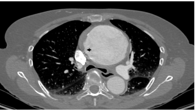
Incomplete dissection with some degree of sub-adventitial hematoma on a computed tomography slice. Notice the intimal tear (arrow).

Magnetic resonance images (echo-gradient sequence, axial and sagittal views) showing intramural aortic hematoma. An ulcer-like projection is observed at the abdominal aorta (arrow).

Histological section (hematoxylin eosin staining) of a patient with intramural hematoma. Hematoma (asterisk) within the aortic media is well documented.
References
Publication types
MeSH terms
Substances
LinkOut - more resources
Full Text Sources
