. 2020 Jun 23:12:4889-4898.
doi: 10.2147/CMAR.S241620. eCollection 2020.

CCNY Accelerates Cylcin E Expression to Regulate the Proliferation of Laryngeal Carcinoma Cells via MEK/ERK Signaling Pathway

Affiliations

CCNY Accelerates Cylcin E Expression to Regulate the Proliferation of Laryngeal Carcinoma Cells via MEK/ERK Signaling Pathway

Xiaoting Zhao et al. Cancer Manag Res. .

Abstract

Background: Laryngeal carcinoma is a common cancer among head and neck tumors, accounting for 0.5-1% new cancer cases or deaths of all tumors throughout the body. Despite improvements in diagnostic and therapy, the prognosis of laryngeal carcinoma patients still remains poor. Thus, it is very important to identify the biomarkers involved in the molecular pathogenesis of laryngeal carcinoma. Cyclin Y (CCNY) is a conserved cell cycle regulator that acts as a growth factor in many cancers. The clinical significance of CCNY in laryngeal carcinoma remains unknown. The function of CCNY in laryngocarcinoma was studied in this paper.

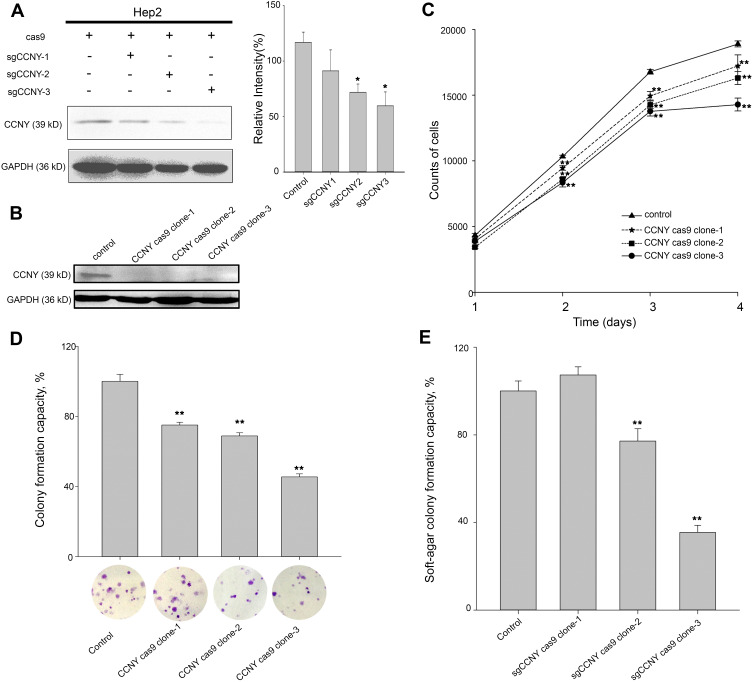
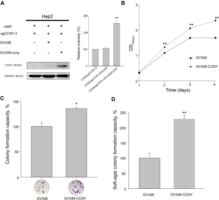
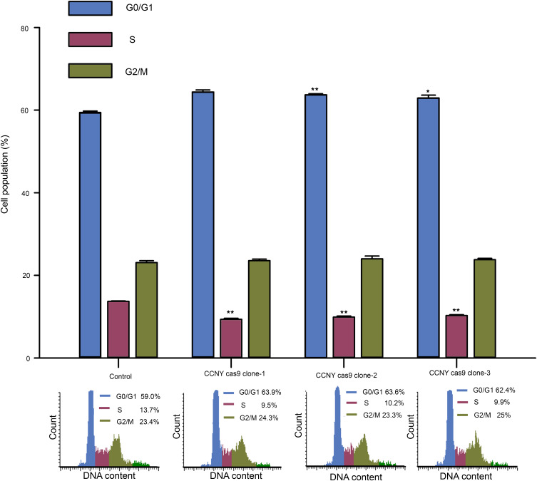
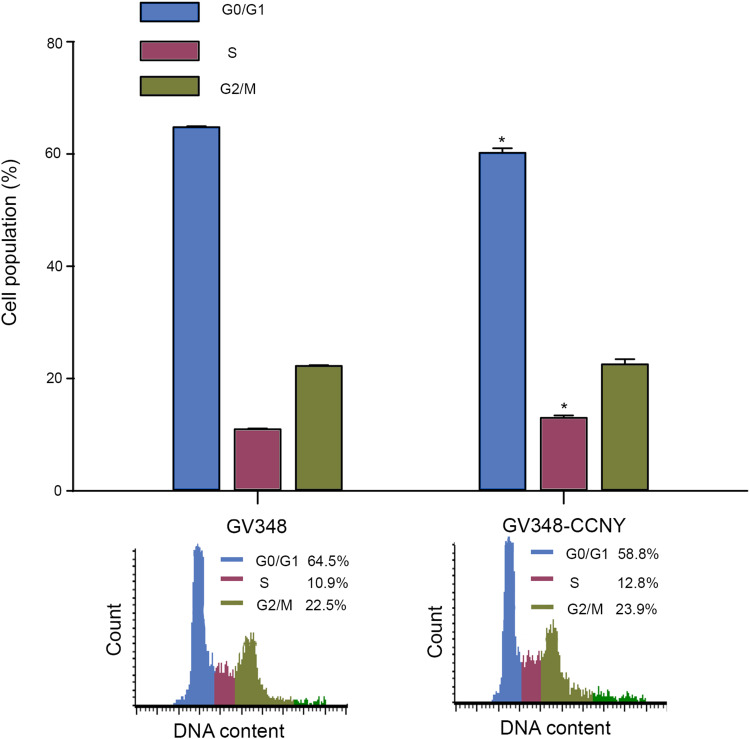
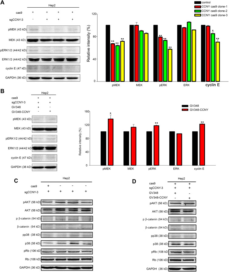
Materials and methods: CCNY knock-out cells were constructed by CRISPR/CAS9 technique. CCNY overexpression cells were also constructed based on CCNY knock-out cells. Cell growth ability was detected by MTS assay, high-content cell analysis, colony formation assays, and anchorage-independent growth assays. The protein levels in laryngocarcinoma cells were determined by Western blot. The role of CCNY in cell cycle progression was evaluated by flow cytometry.

Results: CCNY knock-out cells and CCNY up-regulation cell models were obtained successfully. Suppression of CCNY expression inhibited Hep2 cell growth. Cell growth was enhanced by the up-regulation of CCNY. The percentage of cells in G1 phase was altered when CCNY expression was down-regulated or up-regulated. The phosphorylation level of MEK and ERK as well as cyclin E protein level was also regulated by the expression level of CCNY.

Conclusion: In laryngocarcinoma cell line Hep2 cells, cell proliferation was controlled by CCNY. The expression of CCNY was involved in the cell cycle progress of Hep2 cells. It indicated that CCNY could promote cell growth by activating MEK/ERK/cyclin E signaling pathway.

Keywords: CCNY; ERK; cell cycle; cyclin E; laryngocarcinoma.


Conflict of interest statement

The authors report no conflicts of interest in this work.

Figures

Figure 1
Figure 1
Cell growth ability was suppressed by the knock-out of CCNY. (A) CRISPR/CAS9 technique was used to construct the CCNY knockout cell strains. Three different sgCCNYs (sgCCNY-1, sgCCNY-2, sgCCNY-3) were used. The left panel, the expression level of CCNY was detected by Western blot, with GADPH used as the internal loading controls. The right panel, the level of CCNY was quantified by gray analysis. *P<0.05 (t-test). (B) Three CCNY knock-out cell clones (CCNY cas9 clone-1, CCNY cas9 clone-2, CCNY cas9 clone-3) derived from cells infected by recombinant lentivirus with sgCCNY-3 were identified by Western blot. (C) Cell growth curve was determined by cell counts. The total number of cells were counted by Cellomics ArrayScan HCS Reader. **P<0.01 (t-test). (D) Colony formation efficiency of cells. The data are expressed as the mean ± standard error. **P<0.01 (t-test). (E) Soft-agar colony formation ability of cells. The data are expressed as the mean ± standard error. **P<0.01 (t-test).
Figure 2
Figure 2
Cell proliferation was enhanced by the overexpression of CCNY. (A) CCNY was re-expressed in Hep2 cells by the infection of recombinant lentivirus. The left panel, the expression of CCNY was determined by WB. GAPDH is used as a loading control. The right panel, the level of CCNY was quantified by gray analysis. **P<0.01 (t-test). (B) Cells were subcultured in 96-well plates and an MTS assay was performed. Absorbance at 490 nm (y axis) was determined at every 24-hrs interval. The data are expressed as the mean ± standard error. **P<0.01 (t-test). (C) Colony formation efficiency of cells. The data are expressed as the mean ± standard error. *P<0.05 (t-test). (D) Soft-agar colony formation ability of cells. The data are expressed as the mean ± standard error. **P<0.01 (t-test).
Figure 3
Figure 3
Cell cycle G1/S phase arrest was induced by the suppression of CCNY expression. Cell cycle distribution of cells was determined by flow cytometry. The histogram showed the statistical data expressed as the mean ± standard error. *P<0.05 (t-test), **P<0.01 (t-test).
Figure 4
Figure 4
Cell cycle analysis. Cell cycle distribution of cells was determined by flow cytometry. The histogram showed the statistical data expressed as the mean ± standard error. *P<0.05 (t-test).
Figure 5
Figure 5
CCNY could induce cyclin E expression via MEK/ERK signaling pathway. (A and B) Western blot analysis of ERK1/2, MEK and their phosphorylated forms as well as cyclin E. GAPDH was used as internal loading controls. The levels of proteins were quantified by gray analysis as shown in the right panel. *P<0.05 (t-test), **P<0.01 (t-test). (C and D) The levels of pAKT, AKT, p β-catenin, β-catenin, pp38, p38, pRb, Rb were detected by Western blot. GAPDH was used as internal loading controls.

Similar articles

References

    1. Siegel RL, Miller KD, Jemal A. Cancer statistics, 2019. CA Cancer J Clin. 2019;69(1):7–34. doi:10.3322/caac.21551 - DOI - PubMed
    1. Chen W, Zheng R, Baade PD, et al. Cancer statistics in China, 2015. CA Cancer J Clin. 2016;66(2):115–132. doi:10.3322/caac.21338 - DOI - PubMed
    1. Amemiya K, Shibuya H, Yoshimura R, et al. The risk of radiation-induced cancer in patients with squamous cell carcinoma of the head and neck and its results of treatment. Br J Radiol. 2005;78(935):1028–1033. doi:10.1259/bjr/86352309 - DOI - PubMed
    1. Siddiqui F, Movsas B. Management of radiation toxicity in head and neck cancers. Semin Radiat Oncol. 2017;27(4):340–349. doi:10.1016/j.semradonc.2017.04.008 - DOI - PubMed
    1. Xu W, Han D, Hou L, et al. Voice function following CO2 laser microsurgery for precancerous and early-stage glottic carcinoma. Acta Otolaryngol. 2007;127(6):637–641. doi:10.1080/00016480600987776 - DOI - PubMed