

Assessment of diaphragmatic function by ultrasonography: Current approach and perspectives
- PMID: 32607319
- PMCID: PMC7322428
- DOI: 10.12998/wjcc.v8.i12.2408
Assessment of diaphragmatic function by ultrasonography: Current approach and perspectives
Abstract
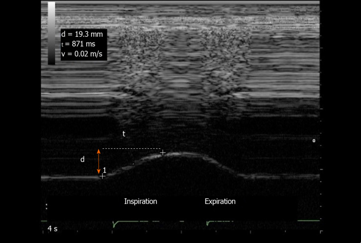
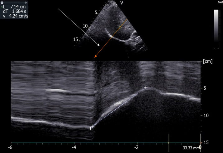
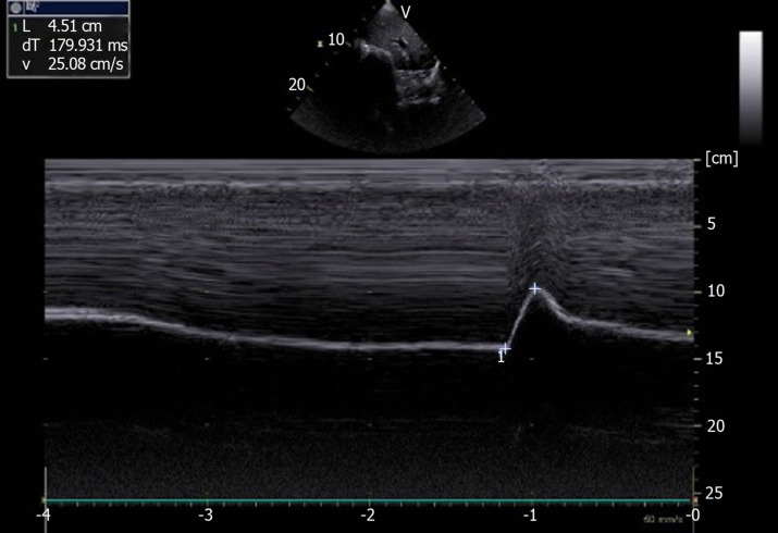
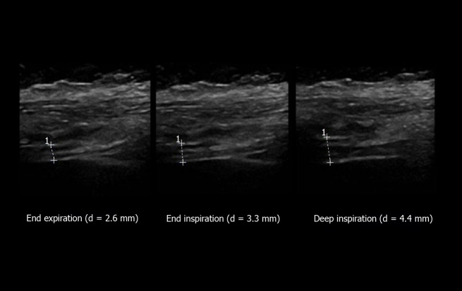
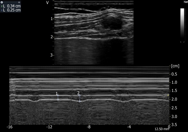
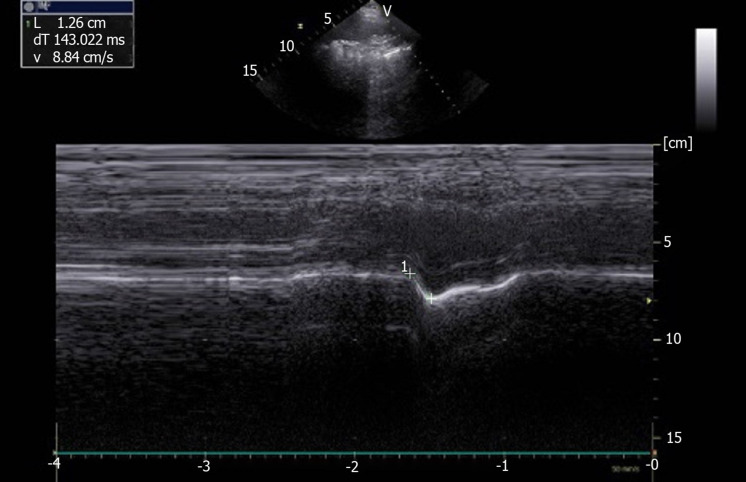
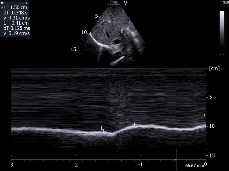
This article reports the various methods used to assess diaphragmatic function by ultrasonography. The excursions of the two hemidiaphragms can be measured using two-dimensional or M-mode ultrasonography, during respiratory maneuvers such as quiet breathing, voluntary sniffing and deep inspiration. On the zone of apposition to the rib cage for both hemidiaphragms, it is possible to measure the thickness on expiration and during deep breathing to assess the percentage of thickening during inspiration. These two approaches make it possible to assess the quality of the diaphragmatic function and the diagnosis of diaphragmatic paralysis or dysfunction. These methods are particularly useful in circumstances where there is a high risk of phrenic nerve injury or in diseases affecting the contractility or the motion of the diaphragm such as neuro-muscular diseases. Recent methods such as speckle tracking imaging and ultrasound shear wave elastography should provide more detailed information for better assessment of diaphragmatic function.
Keywords: Dysfunction; Hemidiaphragm; M-mode; Motion; Paralysis; Speckle tracking imaging; Thickness; Two-dimensional mode; Ultrasound; Ultrasound shear wave elastography.
©The Author(s) 2020. Published by Baishideng Publishing Group Inc. All rights reserved.
Conflict of interest statement
Conflict-of-interest statement: No conflict of interest.
Figures
References
-
- Harris RS, Giovannetti M, Kim BK. Normal ventilatory movement of the right hemidiaphragm studied by ultrasonography and pneumotachography. Radiology. 1983;146:141–144. - PubMed
-
- Houston JG, Morris AD, Howie CA, Reid JL, McMillan N. Technical report: quantitative assessment of diaphragmatic movement--a reproducible method using ultrasound. Clin Radiol. 1992;46:405–407. - PubMed
-
- Targhetta R, Chavagneux R, Ayoub J, Lemerre C, Préfaut C, Bourgeois JM, Balmes P. [Right diaphragmatic kinetics measured by TM-mode ultrasonography with concomitant spirometry in normal subjects and asthmatic patients. Preliminary results] Rev Med Interne. 1995;16:819–826. - PubMed
-
- Kantarci F, Mihmanli I, Demirel MK, Harmanci K, Akman C, Aydogan F, Mihmanli A, Uysal O. Normal diaphragmatic motion and the effects of body composition: determination with M-mode sonography. J Ultrasound Med. 2004;23:255–260. - PubMed
Publication types
LinkOut - more resources
Full Text Sources
Medical
