

Evaluation of internal and shell stiffness in the differential diagnosis of breast non-mass lesions by shear wave elastography
- PMID: 32607328
- PMCID: PMC7322423
- DOI: 10.12998/wjcc.v8.i12.2510
Evaluation of internal and shell stiffness in the differential diagnosis of breast non-mass lesions by shear wave elastography
Abstract
Background: The diagnostic specificity of conventional ultrasound for breast non-mass lesions (NMLs) is low at approximately 21%-43%. Shear wave elastography (SWE) can distinguish benign from malignant lesions by evaluating the internal and peripheral stiffness. SWE has good reproducibility and high diagnostic efficacy. However, there are very few independent studies on the diagnostic value of SWE in breast NMLs.
Aim: To determine the value of SWE in the differential diagnosis of breast NMLs.
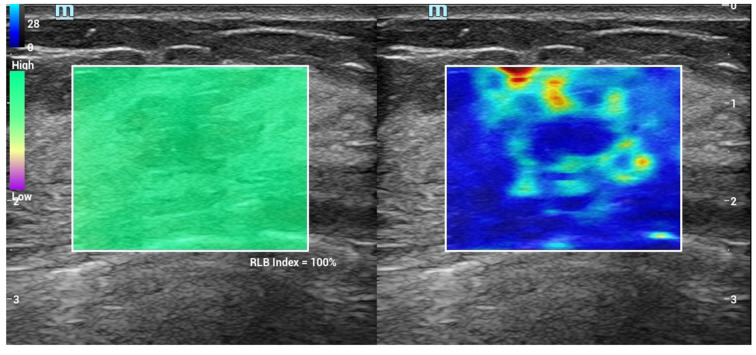
Methods: This study enrolled a total of 118 patients with breast NMLs who underwent SWE examinations in the Beijing Shijitan Hospital Affiliated to Capital Medical University and The Second Hospital of Shandong University from January 2019 to January 2020. The internal elastic parameters of the lesions were recorded, including maximum (Emax), mean (Emean) and minimum elastic values and the standard deviation. The following peripheral parameters were noted: Presence of a "stiff rim" sign; Emax, and Emean elasticity values within 1 mm, 1.5 mm, 2 mm, 2.5 mm and 3 mm from the edge of NMLs. The receiver operating characteristic curve of each parameter was drawn, and the areas under the curve were calculated.
Results: Emax, Emean and elastic values, and the standard deviation of the internal elastic values in malignant NMLs were significantly higher than those in benign NMLs (P < 0.05). The percentage with the "stiff rim" sign in malignant NMLs was significantly higher than that in the benign group (P < 0.05), and Emax and Emean at the shell of 1 mm, 1.5 mm, 2 mm, 2.5 mm and 3 mm in the malignant group were all higher than those in the benign group (P < 0.05). Of the surrounding elasticity values, Emax of the shell at 2.5 mm in malignant NMLs had maximum areas under the curve of 0.900, and the corresponding sensitivity and specificity were 94.57% and 85.86%, respectively.
Conclusion: The "stiff rim" sign and multiple quantitative elastic values within and around the lesion had good diagnostic performance in the differential diagnosis of breast NMLs. Emax in peripheral tissue had better diagnostic efficiency than other parameters.
Keywords: Breast tumor; Diagnosis; Non-mass lesions; Shear wave elastography; Stiff rim sign; Ultrasound.
©The Author(s) 2020. Published by Baishideng Publishing Group Inc. All rights reserved.
Conflict of interest statement
Conflict-of-interest statement: All authors declare no conflicts-of-interest related to this article.
Figures


Similar articles
-
Can measuring perilesional tissue stiffness and stiff rim sign improve the diagnostic performance between benign and malignant breast lesions?J Med Ultrason (2001). 2021 Jan;48(1):53-61. doi: 10.1007/s10396-020-01064-0. Epub 2021 Jan 4. J Med Ultrason (2001). 2021. PMID: 33439373
-
The role of tissue elasticity in the differential diagnosis of benign and malignant breast lesions using shear wave elastography.BMC Cancer. 2020 Sep 29;20(1):930. doi: 10.1186/s12885-020-07423-x. BMC Cancer. 2020. PMID: 32993571 Free PMC article.
-
Shear wave elastography in the diagnosis of breast non-mass lesions: factors associated with false negative and false positive results.Eur Radiol. 2017 Sep;27(9):3788-3798. doi: 10.1007/s00330-017-4763-6. Epub 2017 Feb 6. Eur Radiol. 2017. PMID: 28168373
-
Diagnostic performance of elastography for breast non-mass lesions: A systematic review and meta-analysis.Eur J Radiol. 2021 Nov;144:109991. doi: 10.1016/j.ejrad.2021.109991. Epub 2021 Oct 2. Eur J Radiol. 2021. PMID: 34638081
-
Evaluating Different Quantitative Shear Wave Parameters of Ultrasound Elastography in the Diagnosis of Lymph Node Malignancies: A Systematic Review and Meta-Analysis.Cancers (Basel). 2022 Nov 13;14(22):5568. doi: 10.3390/cancers14225568. Cancers (Basel). 2022. PMID: 36428661 Free PMC article. Review.
Cited by
-
Clinical use and adjustment of ultrasound elastography for breast lesions followed WFUMB guidelines and recommendations in the real world.Front Oncol. 2022 Nov 24;12:1022917. doi: 10.3389/fonc.2022.1022917. eCollection 2022. Front Oncol. 2022. PMID: 36505783 Free PMC article.
-
Can shear wave elastography be utilized as an additional tool for the assessment of non-mass breast lesions?Ultrasound. 2022 Feb;30(1):44-51. doi: 10.1177/1742271X21998721. Epub 2021 Mar 17. Ultrasound. 2022. PMID: 35173778 Free PMC article.
-
Convolutional neural network based on automatic segmentation of peritumoral shear-wave elastography images for predicting breast cancer.Front Oncol. 2023 Feb 14;13:1099650. doi: 10.3389/fonc.2023.1099650. eCollection 2023. Front Oncol. 2023. PMID: 36865812 Free PMC article.
-
Diagnostic accuracy of contrast-enhanced ultrasound synchronized with shear wave elastography in the differential diagnosis of benign and malignant breast lesions: a diagnostic test.Gland Surg. 2023 Jan 1;12(1):54-66. doi: 10.21037/gs-22-684. Epub 2023 Jan 13. Gland Surg. 2023. PMID: 36761482 Free PMC article.
-
Shear wave elastography primer for the abdominal radiologist.Abdom Radiol (NY). 2025 Aug;50(8):3744-3763. doi: 10.1007/s00261-025-04806-1. Epub 2025 Jan 30. Abdom Radiol (NY). 2025. PMID: 39883164 Review.
References
-
- Torre LA, Bray F, Siegel RL, Ferlay J, Lortet-Tieulent J, Jemal A. Global cancer statistics, 2012. CA Cancer J Clin. 2015;65:87–108. - PubMed
-
- Are C, Rajaram S, Are M, Raj H, Anderson BO, Chaluvarya Swamy R, Vijayakumar M, Song T, Pandey M, Edney JA, Cazap EL. A review of global cancer burden: trends, challenges, strategies, and a role for surgeons. J Surg Oncol. 2013;107:221–226. - PubMed
-
- Ko KH, Hsu HH, Yu JC, Peng YJ, Tung HJ, Chu CM, Chang TH, Chang WC, Wu YC, Lin YP, Hsu GC. Non-mass-like breast lesions at ultrasonography: feature analysis and BI-RADS assessment. Eur J Radiol. 2015;84:77–85. - PubMed
-
- Wang ZL, Li N, Li M, Wan WB. Non-mass-like lesions on breast ultrasound: classification and correlation with histology. Radiol Med. 2015;120:905–910. - PubMed
-
- Choi JS, Han BK, Ko EY, Ko ES, Shin JH, Kim GR. Additional diagnostic value of shear-wave elastography and color Doppler US for evaluation of breast non-mass lesions detected at B-mode US. Eur Radiol. 2016;26:3542–3549. - PubMed
LinkOut - more resources
Full Text Sources
