

Connectome mapping with edge density imaging differentiates pediatric mild traumatic brain injury from typically developing controls: proof of concept
- PMID: 32607611
- PMCID: PMC7501221
- DOI: 10.1007/s00247-020-04743-9
Connectome mapping with edge density imaging differentiates pediatric mild traumatic brain injury from typically developing controls: proof of concept
Abstract
Background: Although acute neurologic impairment might be transient, other long-term effects can be observed with mild traumatic brain injury. However, when pediatric patients with mild traumatic brain injury present for medical care, conventional imaging with CT and MR imaging often does not reveal abnormalities.
Objective: To determine whether edge density imaging can separate pediatric mild traumatic brain injury from typically developing controls.
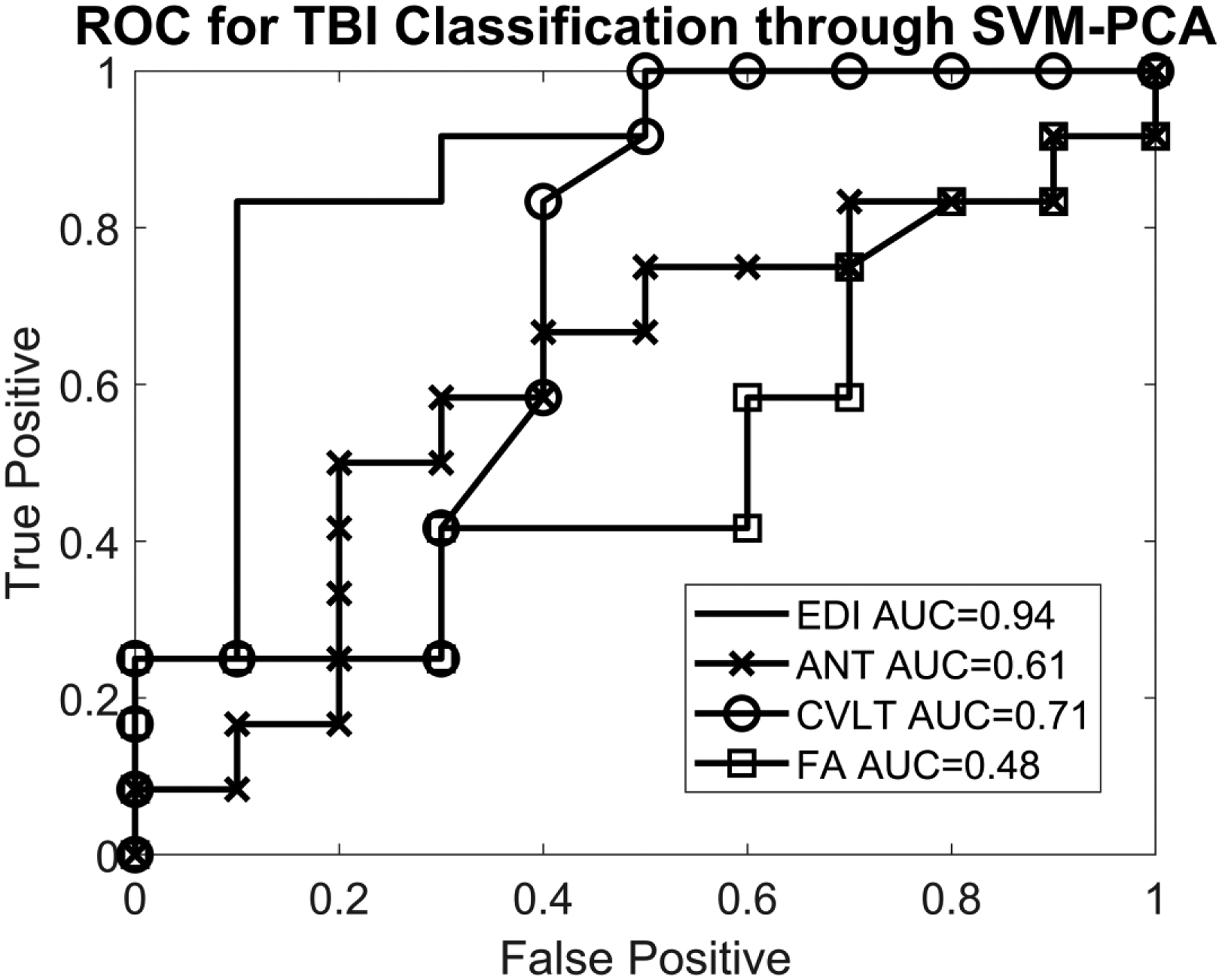
Materials and methods: Subjects were recruited as part of the "Therapeutic Resources for Attention Improvement using Neuroimaging in Traumatic Brain Injury" (TRAIN-TBI) study. We included 24 adolescents (χ=14.1 years of age, σ=1.6 years, range 10-16 years), 14 with mild traumatic brain injury (TBI) and 10 typically developing controls. Neurocognitive assessments included the pediatric version of the California Verbal Learning Test (CVLT) and the Attention Network Task (ANT). Diffusion MR imaging was acquired on a 3-tesla (T) scanner. Edge density images were computed utilizing fiber tractography. Principal component analysis (PCA) and support vector machines (SVM) were used in an exploratory analysis to separate mild TBI and control groups. The diagnostic accuracy of edge density imaging, neurocognitive tests, and fractional anisotropy (FA) from diffusion tensor imaging (DTI) was computed with two-sample t-tests and receiver operating characteristic (ROC) metrics.
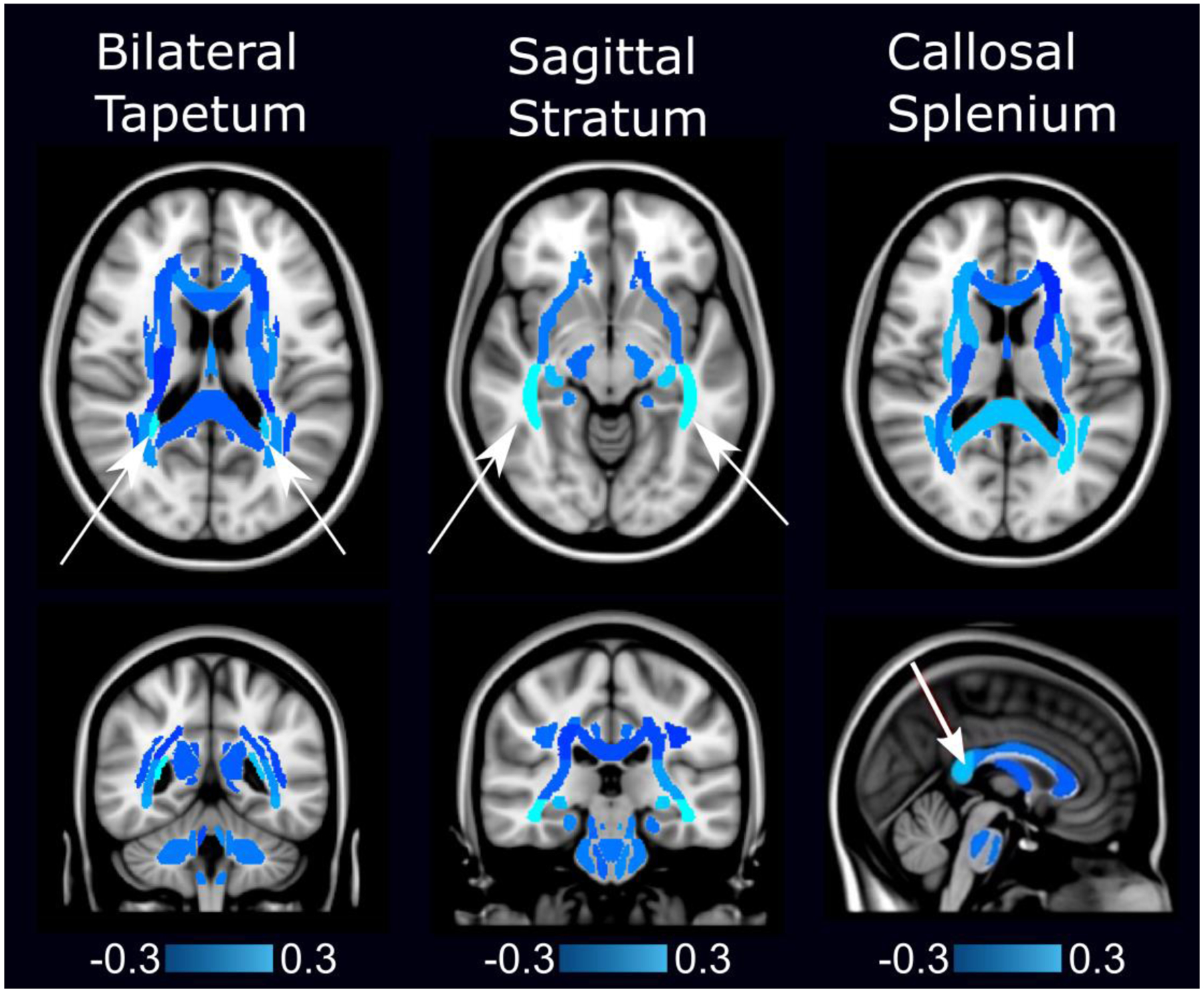
Results: Support vector machine-principal component analysis of edge density imaging maps identified three white matter regions distinguishing pediatric mild TBI from controls. The bilateral tapetum, sagittal stratum, and callosal splenium identified mild TBI subjects with sensitivity of 79% and specificity of 100%. Accuracy from the area under the ROC curve (AUC) was 94%. Neurocognitive testing provided an AUC of 61% (CVLT) and 71% (ANT). Fractional anisotropy yielded an AUC of 48%.
Conclusion: In this proof-of-concept study, we show that edge density imaging is a new form of connectome mapping that provides better diagnostic delineation between pediatric mild TBI and healthy controls than DTI or neurocognitive assessments of memory or attention.
Keywords: Brain; Children; Concussion; Diffusion tensor imaging; Edge density imaging; Magnetic resonance imaging; Traumatic brain injury.
Conflict of interest statement
Figures

Similar articles
-
Relevance of neuroimaging for neurocognitive and behavioral outcome after pediatric traumatic brain injury.Brain Imaging Behav. 2018 Feb;12(1):29-43. doi: 10.1007/s11682-017-9673-3. Brain Imaging Behav. 2018. PMID: 28092022 Free PMC article.
-
White Matter Connectome Edge Density in Children with Autism Spectrum Disorders: Potential Imaging Biomarkers Using Machine-Learning Models.Brain Connect. 2019 Mar;9(2):209-220. doi: 10.1089/brain.2018.0658. Brain Connect. 2019. PMID: 30661372 Free PMC article.
-
Acute pediatric traumatic brain injury severity predicts long-term verbal memory performance through suppression by white matter integrity on diffusion tensor imaging.Brain Imaging Behav. 2020 Oct;14(5):1626-1637. doi: 10.1007/s11682-019-00093-9. Brain Imaging Behav. 2020. PMID: 31134584 Free PMC article.
-
Advanced neuroimaging in traumatic brain injury: an overview.Neurosurg Focus. 2019 Dec 1;47(6):E17. doi: 10.3171/2019.9.FOCUS19652. Neurosurg Focus. 2019. PMID: 32364704 Review.
-
Neuroimaging of structural pathology and connectomics in traumatic brain injury: Toward personalized outcome prediction.Neuroimage Clin. 2012 Aug 24;1(1):1-17. doi: 10.1016/j.nicl.2012.08.002. eCollection 2012. Neuroimage Clin. 2012. PMID: 24179732 Free PMC article. Review.
Cited by
-
MaPPeRTrac: A Massively Parallel, Portable, and Reproducible Tractography Pipeline.Neuroinformatics. 2024 Apr;22(2):177-191. doi: 10.1007/s12021-024-09650-0. Epub 2024 Mar 6. Neuroinformatics. 2024. PMID: 38446357
-
Changes in the Networks of Attention across the Lifespan: A Graphical Meta-Analysis.J Intell. 2024 Feb 10;12(2):19. doi: 10.3390/jintelligence12020019. J Intell. 2024. PMID: 38392175 Free PMC article. Review.
-
Safeguarding Athletes Against Head Injuries Through Advances in Technology: A Scoping Review of the Uses of Machine Learning in the Management of Sports-Related Concussion.Front Sports Act Living. 2022 Apr 20;4:837643. doi: 10.3389/fspor.2022.837643. eCollection 2022. Front Sports Act Living. 2022. PMID: 35520095 Free PMC article.
-
Classification accuracy of structural and functional connectomes across different depressive phenotypes.Imaging Neurosci (Camb). 2024 Jan 17;2:imag_2_00064. doi: 10.1162/imag_a_00064. eCollection 2024. Imaging Neurosci (Camb). 2024. PMID: 40800531 Free PMC article.
-
Structural connectome differences in pediatric mild traumatic brain and orthopedic injury.Hum Brain Mapp. 2022 Feb 15;43(3):1032-1046. doi: 10.1002/hbm.25705. Epub 2021 Nov 8. Hum Brain Mapp. 2022. PMID: 34748258 Free PMC article.
References
-
- Dewan MC, Mummareddy N, Wellons JC, Bonfield CM (2016) Epidemiology of global pediatric traumatic brain injury: qualitative review. World Neurosurg 91:497–509.e1 - PubMed
-
- Pfister T, Pfister K, Hagel B et al. (2016) The incidence of concussion in youth sports: a systematic review and meta-analysis. Br J Sports Med 50:292–297 - PubMed
-
- Kay T, Harrington DE, Adams R et al. (1993) Definition of mild traumatic brain injury. J Head Trauma Rehabil 8:86–87
-
- Guo X, Edmed SL, Anderson V, Kenardy J (2017) Neurocognitive predictors of posttraumatic stress disorder symptoms in children 6 months after traumatic brain injury: a prospective study. Neuropsychology 31:84–92 - PubMed
