. 2020 Sep;94(9):3105-3123.
doi: 10.1007/s00204-020-02826-y. Epub 2020 Jun 30.

Design and evaluation of bi-functional iron chelators for protection of dopaminergic neurons from toxicants

Affiliations

Design and evaluation of bi-functional iron chelators for protection of dopaminergic neurons from toxicants

Simon Gutbier et al. Arch Toxicol. 2020 Sep.

Abstract

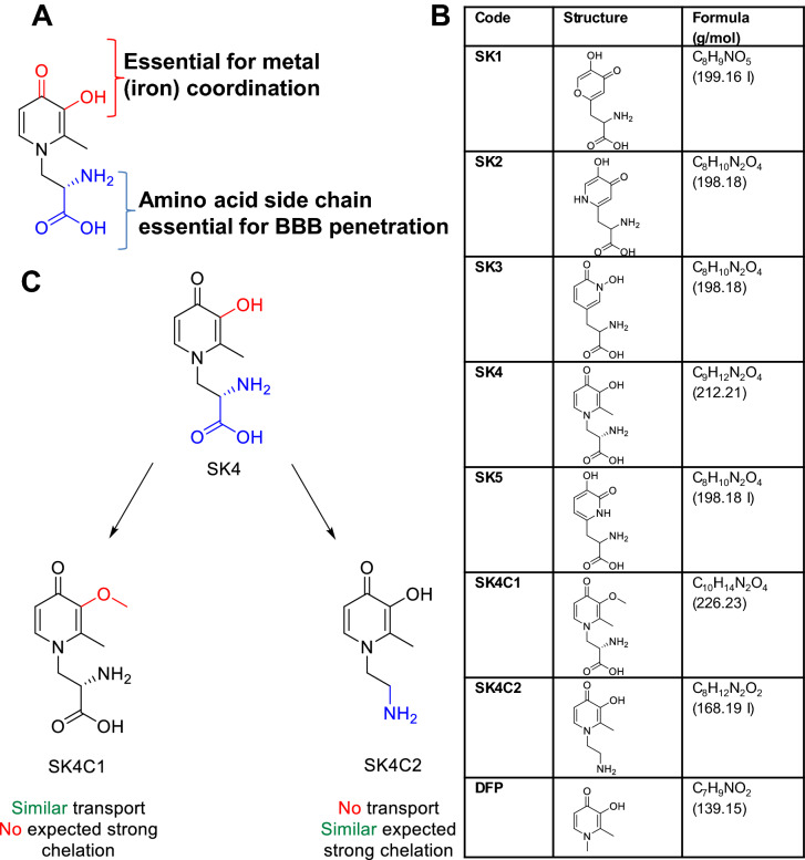
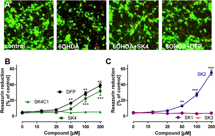
While the etiology of non-familial Parkinson's disease (PD) remains unclear, there is evidence that increased levels of tissue iron may be a contributing factor. Moreover, exposure to some environmental toxicants is considered an additional risk factor. Therefore, brain-targeted iron chelators are of interest as antidotes for poisoning with dopaminergic toxicants, and as potential treatment of PD. We, therefore, designed a series of small molecules with high affinity for ferric iron and containing structural elements to allow their transport to the brain via the neutral amino acid transporter, LAT1 (SLC7A5). Five candidate molecules were synthesized and initially characterized for protection from ferroptosis in human neurons. The promising hydroxypyridinone SK4 was characterized further. Selective iron chelation within the physiological range of pH values and uptake by LAT1 were confirmed. Concentrations of 10-20 µM blocked neurite loss and cell demise triggered by the parkinsonian neurotoxicants, methyl-phenyl-pyridinium (MPP+) and 6-hydroxydopamine (6-OHDA) in human dopaminergic neuronal cultures (LUHMES cells). Rescue was also observed when chelators were given after the toxicant. SK4 derivatives that either lacked LAT1 affinity or had reduced iron chelation potency showed altered activity in our assay panel, as expected. Thus, an iron chelator was developed that revealed neuroprotective properties, as assessed in several models. The data strongly support the role of iron in dopaminergic neurotoxicity and suggests further exploration of the proposed design strategy for improving brain iron chelation.

Keywords: Blood–brain barrier; Dopaminergic neurons; Drug design; Hydroxypyridinones; Iron chelators; LAT1; Parkinson’s disease.


Conflict of interest statement

The authors declare no conflicts of interest.

Figures

Fig. 1
Fig. 1
Design hypothesis for novel iron chelators. Iron chelators characterized in this study were designed for both optimized iron co-ordination and enhanced blood brain barrier transport. a All compounds have two substructures with different function: (i) a group essential for the iron co-ordination (red) and (ii) the amino acid side chain to facilitate LAT1-mediated uptake into endothelial cells of the BBB (blue). b Acronyms, structures and molecular weights of all chelators tested within this study. c Design of proof-of-concept control compounds. SK4C1 and SK4C2 are derivatives of SK4 with a functional inactivation of one of the substructures depicted in a. SK4C1 has a methoxy group (in place of the hydroxy), which inhibits iron chelation and SK4C2 is a primary amine (not an amino acid), which is not transported by LAT1 (color figure online)
Fig. 2
Fig. 2
Protection of neurons from erastin-induced ferroptosis by SK compounds. a Differentiated LUHMES neurons were treated on day 6 (d6) with erastin (1.25 μM) in the presence or absence of SK4 (200 μM) or deferiprone (DFP; 200 μM). After 24 h, the cells were stained with calcein-AM (green stain of live cells) and H-33342 (red stain for live and dead cells). Representative images are shown. The width of one image corresponds to 300 μm of the original cultures b/c: Neuroprotection against erastin-induced ferroptosis of LUHMES (d6) cells was tested as in a, but resazurin reduction was used as quantitative endpoint. D/E: Experiments were performed as described in ac, but neurite integrity was quantified by automated microscopy as alternative endpoint f/g: Experiments were performed as described in ae, but the release of lactate dehydrogenase (LDH) was measured as death endpoint. Data are means + SEM of three independent experiments (color figure online)
Fig. 3
Fig. 3
Antioxidant properties of iron chelators. a Interaction of SK compounds with superoxide. The SK compounds (50 µM) were incubated with xanthine oxidase (1 mU/ml), hypoxanthine (500 µM), and DHE (5 µM) for 20 min. b Interaction of SK compounds (50 µM) with nitric oxide was investigated by their incubation with the ·NO-donor Spermine-NONOate (10 µM) and the detection of free ·NO by a ·NO-selective electrode. As positive control, superoxide was generated by xanthine oxidase/hypoxanthine to quench ·NO. c + d Interaction of SK compounds with peroxynitrite was investigated by application of the peroxynitrite-generating compound Sin-1 (50 µM). As readout, DHR 123 (1 µM) was added, its oxidation was followed by the detection of Rhodamine fluorescence. Desferoxamine and deferriprone (DFP) were tested as alternative iron chelators, uric acid and ascorbic acid served as positive controls. e + f Interference of SK compounds with Fe2+/H2O2-derived hydroxyl radical (·OH) generation. Ferrous iron (20 µM) and H2O2 (50 µM) were combined to allow ·OH generation. As readout, · OH-dependent formation of malondialdehyde was assessed. Data are means + SEM of three independent experiments. Significance tests were not performed for individual data points
Fig. 4
Fig. 4
SK4 transport by LAT1. The transport of SK4 and SK4C2 by LAT1 was assessed in HEK293 cells transfected with human LAT1. Radiolabeled 3[H]-phenylalanine was used as established LAT1 substrate. a Cells were exposed to 3[H]-phenylalanine for 3 min in the presence or absence of competitors (1 mM). Then, the cells were washed, lysed and analyzed for their radioactive content. Uptake was normalized to the data obtained in the absence of competitors. b In a trans-stimulation assay, HEK293 LAT1 cells were preloaded with 3[H]-phenylalanine. Then, they were incubated in the presence of potential LAT1 substrates. The known substrate Leu was used as positive control of a compound that drives the antiporter transport cycle and, thus, accelerates the emptying of cells of LAT1 substrates such as phenylalanine. The cell emptying capacity via LAT1 was then compared for SK4 and SK4C2. Data are expressed as mean ± SD (n = 3) of three independent experiments performed in triplicate. Significantly different from indicated condition: *(P < 0.05), **(P < 0.01), ***(P < 0.001), ****(P < 0.0001)
Fig. 5
Fig. 5
Iron chelating properties of SK4. a Chelation power of ligands for the studied metals (combinations specified in the graph) between pH 5.5 and 8. The y-axis shows the concentration of metal non-complexed to the ligand at the given conditions (− log[Mn+]nc, [L]tot = 0·M, ([M]tot  = 1·M), taking into account the experimentally determined protonation constants of the ligands, the stability constants of their metal complexes, and the hydrolysis constants of the metals. Thus, a value of 6 indicates no complexation by the given ligand (i.e., a free concentration of the metal ion of 1 µM), while a value of 12 indicates that 1 pM is non-complexed. Thus, the higher the value, the stronger the chelator. Note that the line for the SK2-Fe3+ complex is not shown in the diagram, as it runs continuously at 6 (no complex formation in the pH range shown). b Protection of neuronal cells from iron toxicity. Human dopaminergic neurons (LUHMES) were incubated with FeSO4 (150 µM) and SK2 or SK4 at the concentrations indicated for a period of 4 days. For visualization of cell morphology, cells were stained with an anti-β-III-tubulin antibody. Control cells showed a typical neuronal network with many fine neurite processes between the cells. Fe2+ led to cell loss and clumping of the remaining cells. This was prevented by SK4, but not SK2. The width of images corresponds to 200 μm in original cultures. c In experiments performed as in b, viability was assessed by the analysis of resazurin reduction. Data are means ± D of 3 independent experiments. There was a highly significant statistical difference at 100 µM (P < 0.001; ANOVA with Dunnett’s post hoc test)
Fig. 6
Fig. 6
Protection of neurons from MPP+ toxicity by SK compounds. a Differentiated LUHMES neurons were treated on day 6 (d6) with MPP+ (5 μM) in the presence or absence of SK4 (200 μM) or DFP (200 μM). After 72 h, the cells were stained with calcein-AM (green stain of live cells) and H-33342 (red stain for live and dead cells). Representative images are shown. The width of one image corresponds to 300 μm of the original cultures. b/c Experiments were performed as described in a, but neurite integrity was quantified by automated microscopy as alternative endpoint d/e Experiments were performed as described in ac, but the release of lactate dehydrogenase (LDH) was measured as death endpoint. Data are means + SEM of three independent experiments
Fig. 7
Fig. 7
Protection of neurons from 6-hydroxydopamine toxicity by SK compounds. a Differentiated LUHMES neurons were treated on day 6 (d6) with 6-OHDA (100 μM) in the presence or absence of SK4 (200 μM) or deferiprone (DFP; 200 μM). After 18 h, the cells were stained with calcein-AM (green stain of live cells) and H-33342 (red stain for live and dead cells). Representative images are shown. The width of one image corresponds 300 μm of the original cultures b/c Neuroprotection against 6-OHDA-induced neuronal damage of LUHMES (d6) cells was tested as in a, but resazurin reduction was used as quantitative endpoint. Data are means + SEM of three independent experiments
Fig. 8
Fig. 8
Protection by SK4 from erastin-induced ferroptosis in neuronal organoids. a Generation of LUHMES organoids: LUHMES (pre-differentiated for 2 days) were left to self-aggregate (d2–d8), and the resultant spheres were then plated. After 48 h of neurite outgrowth from the organoids (d8–d10) they were treated with erastin (10 μM) for 24 h. Optionally, they were preincubated for 1 h with ferrostatin-1, or the iron chelators desferoxamine (DFO), or SK4. Finally, organoids were stained with calcein-AM and propidium iodide to assess survival endpoints. b Workflow (and exemplary primary and processed images) to quantify the total neurite area (calcein staining) and the cell viability (propidium iodide (PI) fluorescence). c The plated organoids were treated for 24 h with either erastin alone (10 μM) or together with ferrostatin-1 (100 nM), desferoxamine (5 μM) or SK4 (100 μM). Data are means ± SD (three independent experiments with at least three technical replicates each). Statistical significance was determined by one-way ANOVA with Dunnet‘s post hoc test in comparison to erastin treatment. ***P < 0.001. d Representative images of neurite areas stained with PI and calcein under various conditions. e Organoids were fixed and immunostained for neurofilament heavy chains (NF200). Images were recorded by confocal microscopy. Representative images are shown for cultures exposed to erastin in the presence or absence of SK4

Similar articles

Cited by

References

    1. Andersen JK. Oxidative stress in neurodegeneration: cause or consequence? Nat Med. 2004;10(Suppl):S18–25. doi: 10.1038/nrn1434. - DOI - PubMed
    1. Baes CF, Mesmer RE. The hydrolysis of cations. Krieger, Malabar, Fla: R.E; 1986.
    1. Bartesaghi S, Trujillo M, Denicola A, Folkes L, Wardman P, Radi R. Reactions of desferrioxamine with peroxynitrite-derived carbonate and nitrogen dioxide radicals. Free Radic Biol Med. 2004;36(4):471–483. doi: 10.1016/j.freeradbiomed.2003.10.011. - DOI - PubMed
    1. Blum D, Torch S, Lambeng N, et al. Molecular pathways involved in the neurotoxicity of 6-OHDA, dopamine and MPTP: contribution to the apoptotic theory in Parkinson's disease. Prog Neurobiol. 2001;65(2):135–172. doi: 10.1016/s0301-0082(01)00003-x. - DOI - PubMed
    1. Boddaert N, Le Quan Sang KH, Rotig A, et al. Selective iron chelation in Friedreich ataxia: biologic and clinical implications. Blood. 2007;110(1):401–408. doi: 10.1182/blood-2006-12-065433. - DOI - PubMed

Publication types

MeSH terms