

Performance assessment of the 2 γpositronium imaging with the total-body PET scanners
- PMID: 32607664
- PMCID: PMC7326848
- DOI: 10.1186/s40658-020-00307-w
Performance assessment of the 2 γpositronium imaging with the total-body PET scanners
Abstract
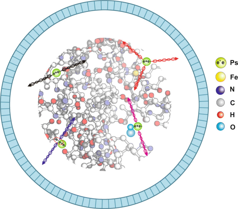
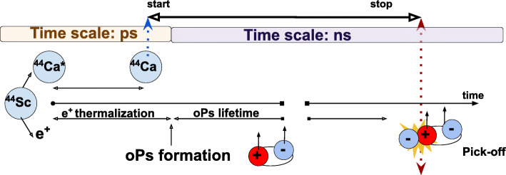
Purpose: In living organisms, the positron-electron annihilation (occurring during the PET imaging) proceeds in about 30% via creation of a metastable ortho-positronium atom. In the tissue, due to the pick-off and conversion processes, over 98% of ortho-positronia annihilate into two 511 keV photons. In this article, we assess the feasibility for reconstruction of the mean ortho-positronium lifetime image based on annihilations into two photons. The main objectives of this work include the (i) estimation of the sensitivity of the total-body PET scanners for the ortho-positronium mean lifetime imaging using 2γ annihilations and (ii) estimation of the spatial and time resolution of the ortho-positronium image as a function of the coincidence resolving time (CRT) of the scanner.
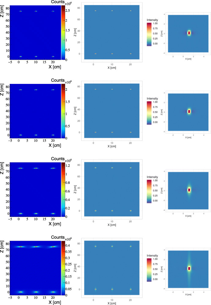
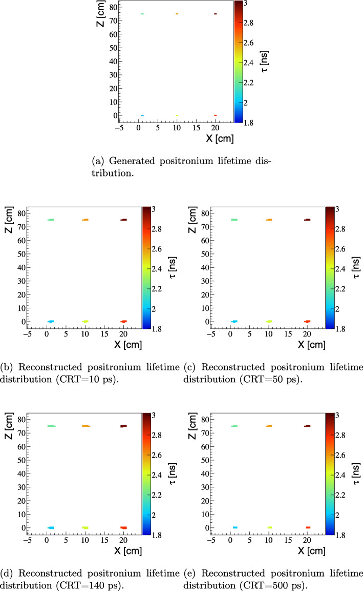
Methods: Simulations are conducted assuming that radiopharmaceutical is labeled with 44Sc isotope emitting one positron and one prompt gamma. The image is reconstructed on the basis of triple coincidence events. The ortho-positronium lifetime spectrum is determined for each voxel of the image. Calculations were performed for cases of total-body detectors build of (i) LYSO scintillators as used in the EXPLORER PET and (ii) plastic scintillators as anticipated for the cost-effective total-body J-PET scanner. To assess the spatial and time resolution, the four cases were considered assuming that CRT is equal to 500 ps, 140 ps, 50 ps, and 10 ps.
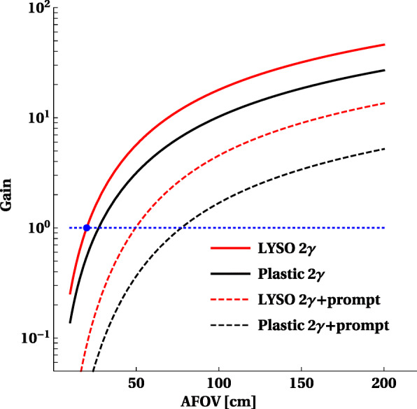
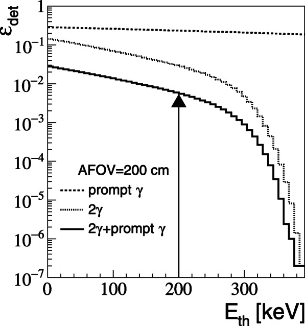
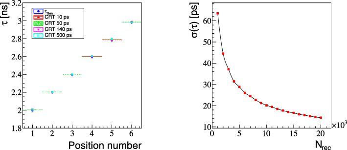
Results: The estimated total-body PET sensitivity for the registration and selection of image forming triple coincidences (2γ+γprompt) is larger by a factor of 13.5 (for LYSO PET) and by factor of 5.2 (for plastic PET) with respect to the sensitivity for the standard 2γ imaging by LYSO PET scanners with AFOV = 20 cm. The spatial resolution of the ortho-positronium image is comparable with the resolution achievable when using TOF-FBP algorithms already for CRT = 50 ps. For the 20-min scan, the resolution better than 20 ps is expected for the mean ortho-positronium lifetime image determination.
Conclusions: Ortho-positronium mean lifetime imaging based on the annihilations into two photons and prompt gamma is shown to be feasible with the advent of the high sensitivity total-body PET systems and time resolution of the order of tens of picoseconds.
Keywords: Medical imaging; PET; Positronium imaging; Total-body PET.
Conflict of interest statement
The authors declare that they have no competing interests.
Figures
References
-
- Moskal P, et al. Feasibility study of the positronium imaging with the J-PET tomograph. Phys Med Biol. 2019;64(5):055017. - PubMed
-
- Harpen MD. Positronium: Review of symmetry, conserved quantities and decay for the radiological physicist. Med Phys. 2004;31(1):57–61. - PubMed
-
- Chen H, Van Horn J, Ching JY. Applications of positron annihilation spectroscopy to life science. 2012; 331:275–93. 10.4028/www.scientific.net/ddf.331.275.
-
- Jasińska B, et al. Human tissues investigation using PALS technique. Acta Phys Polon. 2017;B48:1737.
-
- Pietrzak R, Borbulak S, Szatanik R. Influence of neoplastic therapy on the investigated blood using positron annihilation lifetime spectroscopy. Nukleonika. 2013;58:199–202.
LinkOut - more resources
Full Text Sources
Research Materials
Miscellaneous
