. 2020 Aug;53(8):e12830.
doi: 10.1111/cpr.12830. Epub 2020 Jul 1.

Exosomes released from educated mesenchymal stem cells accelerate cutaneous wound healing via promoting angiogenesis

Affiliations

Exosomes released from educated mesenchymal stem cells accelerate cutaneous wound healing via promoting angiogenesis

Xinyu Qiu et al. Cell Prolif. 2020 Aug.

Abstract

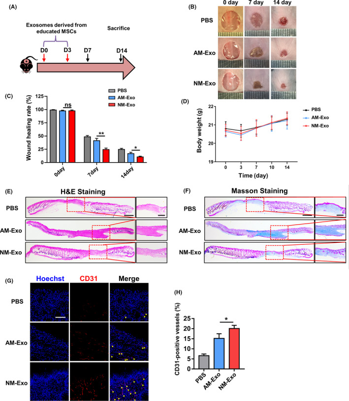
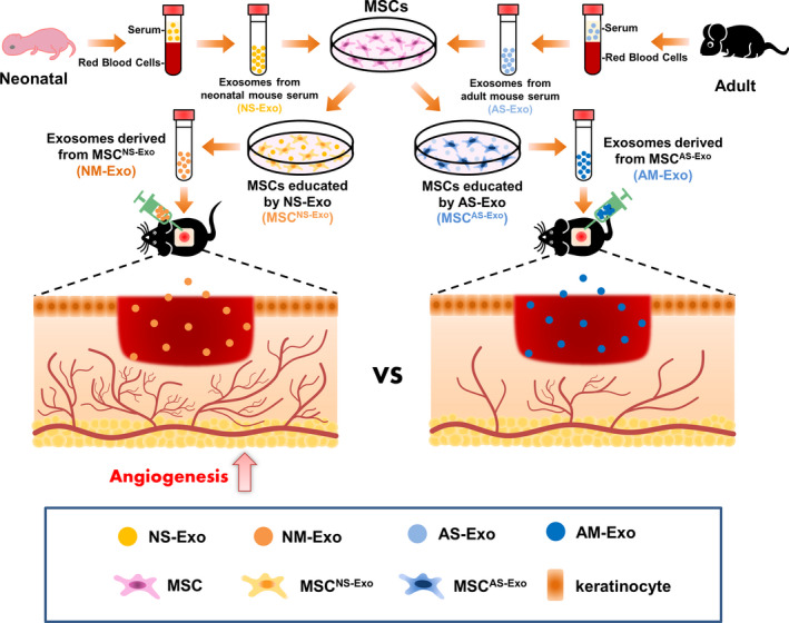
Objectives: Skin serves as the major interface between the external environment and body which is liable to many kinds of injuries. Mesenchymal stem cell (MSC) therapy has been widely used and became a promising strategy. Pre-treatment with chemical agents, hypoxia or gene modifications can partially protect MSCs against injury, and the pre-treated MSCs show the improved differentiation, homing capacity, survival and paracrine effects regard to attenuating injury. The aim of this study was to investigate whether the exosomes from the educated MSCs contribute to accelerate wound healing process.

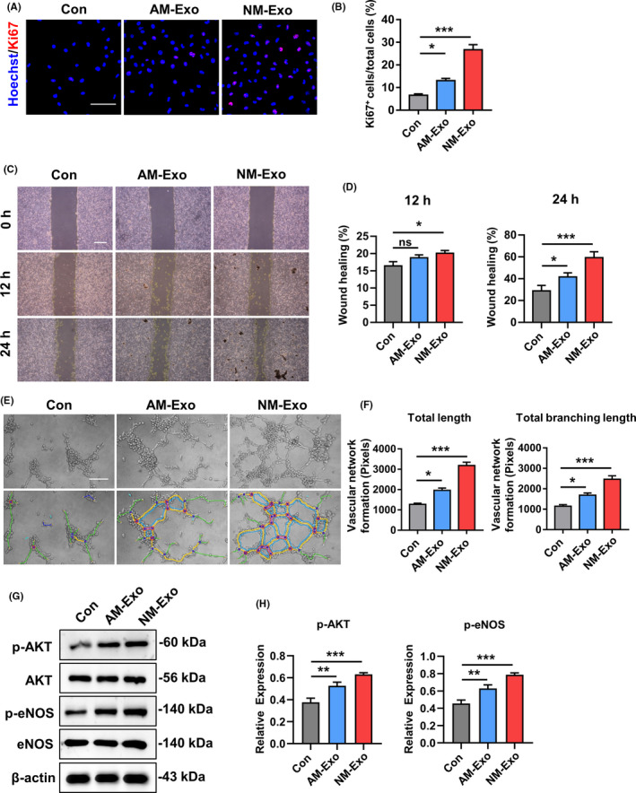
Materials and methods: We extracted the exosomes from the two educated MSCs and utilized them in the cutaneous wound healing model. The pro-angiogenetic effect of exosomes on endothelial cells was also investigated.

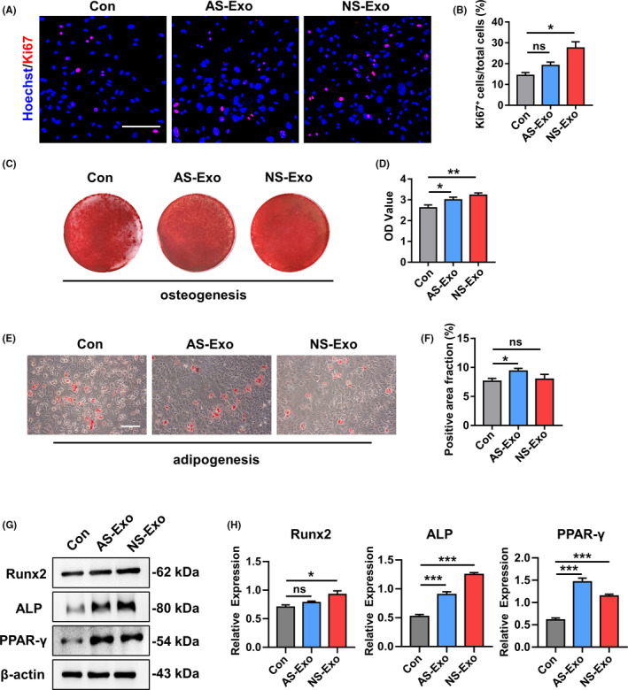
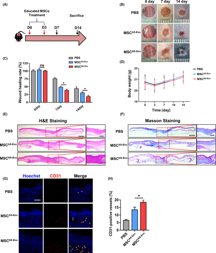
Results: We firstly found that MSCs pre-treated by exosomes from neonatal serum significantly improved their biological functions and the effect of therapy. Moreover, we extracted the exosomes from the educated MSCs and utilized them to treat the cutaneous wound model directly. We found that the released exosomes from MSCs which educated by neonatal serum before had the more outstanding performance in therapeutic effect. Mechanistically, we revealed that the recipient endothelial cells (ECs) were targeted and the exosomes promoted their functions to enhance angiogenesis via regulating AKT/eNOS pathway.

Conclusions: Our findings unravelled the positive effect of the upgraded exosomes from the educated MSCs as a promising cell-free therapeutic strategy for cutaneous wound healing.

Keywords: angiogenesis; exosome; mesenchymal stem cell; regenerative medicine; wound healing.


Conflict of interest statement

The authors have no conflicts of interest to declare.

Figures

FIGURE 1
FIGURE 1
Characterization of bone marrow mesenchymal stem cells (BMMSCs). A, Flow cytometric analysis of ex vivo expanded MSCs revealed positive expression of CD29, CD90, CD105, CD146 and Sca‐1, and negative expression of CD34, CD11b and CD45. B, Representative percentage of cell surface markers for identification. C, Representative proliferation of single clone of bone marrow mesenchymal stem cells. D, MSCs seeded in plates induced with osteogenic medium for 10 days. Activity of ALP detected by ALP staining. E, MSCs cultured in osteogenic inductive conditions for 28 days, mineralized nodules found by Alizarin Red staining. F, Cultured MSCs formed Oil Red O‐positive lipid cluster following 14 days of adipogenic induction. Scale bar, 500 μm. ALP, alkaline phosphatase; APC, allophycocyanin; CFU‐F, fibroblastic colony‐forming unit; FITC, fluorescein isothiocyanate
FIGURE 2
FIGURE 2
Exosome characterization in neonatal and adult serum. A, Transmission electron microscopy showing extracellular vesicles with ~100 nm diameters isolated from neonatal and adult serum. Scale bar, 100 nm. B, Size analysis revealed neonatal serum vesicles with diameters of 109.5 ± 2.1 nm and adult serum vesicles with diameters of 91.3 ± 2.3 nm. C, Total proteins extracted from nanometre vesicles probed by anti‐CD9, anti‐CD63, anti‐CD81 and anti‐TSG101 antibodies. D, The immunofluorescence images of time‐dependent uptake of exosomes by MSCs. E, The immunofluorescence images of concentration‐dependent uptake of exosomes by MSCs. n = 3 per group. Scale bar, 20 μm. AS‐Exo, adult serum exosomes; NS‐Exo, neonatal serum exosomes
FIGURE 3
FIGURE 3
The representative images of Ki‐67 staining (A) and quantified by the positive‐stained percentage through ImageJ software (B). Scale bar, 100 μm. (C‐D) Alizarin Red staining was performed to detect mineralized nodules formed in Con, AS‐Exo and NS‐Exo 14 days after osteogenic induction and quantified with a spectrophotometer after dissolving with cetylpyridinium chloride. (E‐F) Lipid droplet formation was detected by Oil Red O staining 7 days after adipogenic induction with positive area quantified. Scale bar, 100 μm. (G‐H) The protein expression levels of Runx2, ALP and PPAR‐γ in Con, AS‐Exo and NS‐Exo groups were measured through Western blot and quantified by ImageJ software. n = 3 per group. Data are shown as mean ± SD; ns, not significant; *P < .05; **P < .01; ***P < .001. AS‐Exo, adult serum exosomes; Con, control; NS‐Exo, neonatal serum exosomes
FIGURE 4
FIGURE 4
The schematic graph (A) shows the protocol of establishment and treatment for cutaneous wound healing model. (B) The representative photographs of wound healing process of different timepoints in different groups. (C) The quantification of wound healing rate. (D) The quantification of body weight in different groups. (E) Representative images of H&E staining of the skin tissue samples. Scale bar, 1 mm in low magnification images and 500 μm in high magnification images. (F) Representative images of the Masson‐trichrome staining of the skin tissue samples. Scale bar, 1 mm in low magnification images and 500 μm in high magnification images. (G‐H) Representative images and quantification of the CD31 expression in the skin tissue samples in different groups. Yellow arrows indicate the positive area. Scale bar, 100 μm. n = 3 per group. Data are shown as mean ± SD; ns, not significant; *P < .05. PBS, phosphate‐buffered saline; MSCNS‐Exo, MSCs educated by exosomes from neonatal serum; MSCAS‐Exo, MSCs educated by exosomes from adult serum
FIGURE 5
FIGURE 5
The schematic graph (A) shows the protocol of establishment and treatment for cutaneous wound healing model. (B) The representative photographs of wound healing process of different timepoints in different groups. (C) The quantification of wound healing rate. (D) The quantification of body weight in different groups. (E) Representative images of H&E staining of the skin tissue samples. Scale bar, 1 mm in low magnification images and 500 μm in high magnification images. (F) Representative images of the Masson‐trichrome staining of the skin tissue samples. Scale bar, 1 mm in low magnification images and 500 μm in high magnification images. (G‐H) Representative images and quantification of the CD31 expression in the skin tissue samples in different groups. Yellow arrows indicate the positive area. Scale bar, 100 μm. n = 3 per group. Data are shown as mean ± SD; ns, not significant; *P < .05; **P < .01. PBS, phosphate buffer saline; NM‐Exo, exosomes derived from MSCNS‐Exo; AM‐Exo, exosomes derived from MSCAS‐Exo
FIGURE 6
FIGURE 6
Exosomes activated the angiogenic capacity of endothelial cells in vitro. (A) Proliferation of endothelial cells was detected by Ki67 staining and quantified by the positive‐stained percentage (B). Scale bar, 100 μm. (C‐D) Representative images and quantification of scratch assay examining the migration ability of endothelial cells treated with different exosomes. Scale bar, 500 μm. (E‐F) Tube formation capacity on the Matrigel and the images were analysed by ImageJ software. Scale bar, 20 μm. (G‐H) The protein expression levels of p‐AKT, AKT, p‐eNOS and eNOS were detected by Western blot in endothelial cells which were treated with different exosomes. The results were quantified by ImageJ software. n = 3 per group. Data are shown as mean ± SD; ns, not significant; *P < .05; **P < .01; ***P < .001. Con, control; NM‐Exo, exosomes derived from MSCNS‐Exo; AM‐Exo, exosomes derived from MSCAS‐Exo
FIGURE 7
FIGURE 7
Schema for exosomes derived from the educated MSCs promoted angiogenesis in cutaneous wound healing via regulating the functions of endothelial cell

References

    1. Gurtner GC, Werner S, Barrandon Y, Longaker MT. Wound repair and regeneration. Nature. 2008;453(7193):314‐321. - PubMed
    1. Hu MS, Maan ZN, Wu JC, et al. Tissue engineering and regenerative repair in wound healing. Ann Biomed Eng. 2014;42(7):1494‐1507. - PMC - PubMed
    1. Lee DE, Ayoub N, Agrawal DK. Mesenchymal stem cells and cutaneous wound healing: novel methods to increase cell delivery and therapeutic efficacy. Stem Cell Res Ther. 2016;7:37. - PMC - PubMed
    1. Qi Y, Jiang D, Sindrilaru A, et al. TSG‐6 released from intradermally injected mesenchymal stem cells accelerates wound healing and reduces tissue fibrosis in murine full‐thickness skin wounds. J Invest Dermatol. 2014;134(2):526‐537. - PubMed
    1. Pourjafar M, Saidijam M, Mansouri K, Ghasemibasir H, Karimi Dermani F, Najafi R. All‐trans retinoic acid preconditioning enhances proliferation, angiogenesis and migration of mesenchymal stem cell in vitro and enhances wound repair in vivo. Cell Prolif. 2017;50(1):e12315. - PMC - PubMed

MeSH terms