Review
. 2020 Jul 1;130(7):3370-3380.
doi: 10.1172/JCI136226.

The molecular biology and immune control of chronic Toxoplasma gondii infection

Review

The molecular biology and immune control of chronic Toxoplasma gondii infection

Xiao-Yu Zhao et al. J Clin Invest. .

Abstract

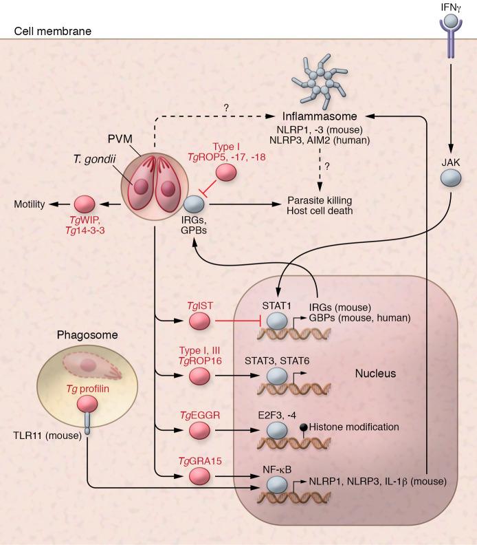
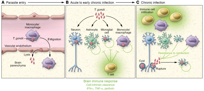
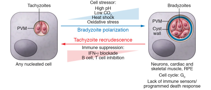
Toxoplasma gondii is an incredibly successful parasite owing in part to its ability to persist within cells for the life of the host. Remarkably, at least 350 host species of T. gondii have been described to date, and it is estimated that 30% of the global human population is chronically infected. The importance of T. gondii in human health was made clear with the first reports of congenital toxoplasmosis in the 1940s. However, the AIDS crisis in the 1980s revealed the prevalence of chronic infection, as patients presented with reactivated chronic toxoplasmosis, underscoring the importance of an intact immune system for parasite control. In the last 40 years, there has been tremendous progress toward understanding the biology of T. gondii infection using rodent models, human cell experimental systems, and clinical data. However, there are still major holes in our understanding of T. gondii biology, including the genes controlling parasite development, the mechanisms of cell-intrinsic immunity to T. gondii in the brain and muscle, and the long-term effects of infection on host homeostasis. The need to better understand the biology of chronic infection is underscored by the recent rise in ocular disease associated with emerging haplotypes of T. gondii and our lack of effective treatments to sterilize chronic infection. This Review discusses the cell types and molecular mediators, both host and parasite, that facilitate persistent T. gondii infection. We highlight the consequences of chronic infection for tissue-specific pathology and identify open questions in this area of host-Toxoplasma interactions.


Conflict of interest statement

Conflict of interest: The authors have declared that no conflict of interest exists.

Figures

Figure 1
Figure 1. Innate immune signaling and the influence of parasite effectors.
T. gondii grows within a parasitophorous vacuole membrane (PVM) that protects the parasite from cytosolic immune sensors and avoids fusion with the endolysosomal compartments containing Toll-like receptors (TLRs). In the mouse, TLR11 recognizes Tg profilin, an actin-modifying protein that is exposed once dead or damaged parasites are phagocytosed. TLR11 is a pseudogene in humans. TgGRA15 can promote host NF-κB phosphorylation and nuclear translocation. In mice, NF-κB stimulation is necessary for transcriptional regulation of the inflammasome components NLRP1, NLRP3, and IL-1; however, human monocytes can engage an NLRP3 inflammasome independent of NF-κB prestimulation. A detailed mechanism of inflammasome activation, parasite killing, and host cell death remains elusive, particularly in regard to signal integration with IFN-γ. IFN-γ signaling induces STAT1 translocation to the nucleus and upregulation of IFN-responsive genes, including immunity-related GTPases (IRGs, mouse) and guanylate-binding proteins (GBPs, human and mouse), which functions to attack parasite vacuole, leading to parasite killing and host cell death. In human cells, GBP1 is necessary for this process, which leads to AIM2 activation of an alternative apoptosis pathway. The type I parasite rhoptry proteins, TgROP5, 17, and 18, can dismantle the function of the mouse IRGs IRGa6 and IRGb6 at the PVM, inactivating GBP attack and parasite killing. The parasite dense granule effector TgIST is a nuclear repressor of STAT1 transcription. TgROP16 is a kinase that phosphorylates and activates host STAT3 and STAT6. TgEGGR affects host gene expression through E2F3- and E2F4-mediated epigenetic modifications. In infected monocytes and dendritic cells (DCs), TgWIP and 14-3-3 proteins promote cell mobility, a putative mechanism of intracellular parasite dissemination in vivo.
Figure 2
Figure 2. T. gondii entry and control of persistent infection in the brain.
(A) In acute infection, T. gondii is frequently observed in immune cells, including monocytes and dendritic cells, with hypermigratory behavior. During infection, blood-brain barrier (BBB) permeability increases and monocytes accumulate in the endothelial lumen, interacting with endothelial cells. These observations have led to the hypothesis that migratory immune cells deliver T. gondii to the BBB and, perhaps, smuggle them into the brain. Replicating parasites are also observed in brain endothelial cells, whose subsequent lysis may be a mechanism of T. gondii entry into the brain. (B) During acute infection parasites are observed infecting neurons, astrocytes, microglia, and infiltrating immune cells. Astrocytes and microglia as well as peripheral monocytes can clear parasites with cell-autonomous immune pathways. (C) As chronic infection progresses, infected astrocytes and microglia or the parasites within them are cleared and cysts are primarily observed within neurons. Most parasite cysts are not associated with immune infiltrate; however, individual parasites or parasite debris can be observed colocalizing with immune infiltrate.
Figure 3
Figure 3. Environmental and host cell–specific pressures driving the T.
gondiitachyzoite to bradyzoite transition. Left: T. gondii tachyzoites can invade almost any nucleated host cell type and grow within the PVM formed from host plasma membrane. In vitro, a range of tissue culture stress conditions can upregulate bradyzoite-specific genes. As parasites polarize to a bradyzoite transcriptional profile, they synthesize a heavily glycosylated cyst wall beneath the PVM. The frequency and rate of bradyzoite differentiation are also influenced by the host cell type, cell cycle status, the host cell lifespan, and inflammatory signals in vitro. In vivo, cysts are most frequently observed in neurons, cardiac muscle, skeletal muscle, and retinal pigment epithelial cells. If the host is immune-suppressed, parasites shift toward a replicative tachyzoite form in a process referred to as recrudescence, which is associated with tissue damage, particularly in the eye.

Similar articles

Cited by

References

    1. Dubey JP, Miller NL, Frenkel JK. The Toxoplasma gondii oocyst from cat feces. J Exp Med. 1970;132(4):636–662. doi: 10.1084/jem.132.4.636. - DOI - PMC - PubMed
    1. Black MW, Boothroyd JC. Lytic cycle of Toxoplasma gondii. Microbiol Mol Biol Rev. 2000;64(3):607–623. doi: 10.1128/MMBR.64.3.607-623.2000. - DOI - PMC - PubMed
    1. Jones JL, Dargelas V, Roberts J, Press C, Remington JS, Montoya JG. Risk factors for Toxoplasma gondii infection in the United States. Clin Infect Dis. 2009;49(6):878–884. doi: 10.1086/605433. - DOI - PubMed
    1. Dubey JP, Speer CA, Shen SK, Kwok OC, Blixt JA. Oocyst-induced murine toxoplasmosis: life cycle, pathogenicity, and stage conversion in mice fed Toxoplasma gondii oocysts. J Parasitol. 1997;83(5):870–882. doi: 10.2307/3284282. - DOI - PubMed
    1. Speer CA, Dubey JP. Ultrastructure of early stages of infections in mice fed Toxoplasma gondii oocysts. Parasitology. 1998;116(pt 1):35–42. - PubMed