

Neurologic and Radiographic Findings Associated With COVID-19 Infection in Children
- PMID: 32609336
- PMCID: PMC7330822
- DOI: 10.1001/jamaneurol.2020.2687
Neurologic and Radiographic Findings Associated With COVID-19 Infection in Children
Erratum in
-
Omitted Related Publication and Acknowledgment.JAMA Neurol. 2020 Dec 1;77(12):1582. doi: 10.1001/jamaneurol.2020.3946. JAMA Neurol. 2020. PMID: 33044506 Free PMC article. No abstract available.
Abstract
Importance: Neurological manifestations have been reported in adults with coronavirus disease 2019 (COVID-19), which is caused by the highly pathogenic virus severe acute respiratory syndrome coronavirus 2 (SARS-CoV-2).
Objective: To report the neurological manifestations of children with COVID-19.
Design, setting, and participants: In this case-series study, patients younger than 18 years who presented with SARS-CoV-2 infection and neurological symptoms to Great Ormond Street Hospital for Children (London, UK) between March 1, 2020, and May 8, 2020, were included after infection was confirmed by either a quantitative reverse transcription-polymerase chain reaction assay by nasopharyngeal swab or a positive test result for IgG antibodies against SARS-CoV-2 in serum.
Main outcomes and measures: Clinical and paraclinical features were retrieved from electronic patient records.
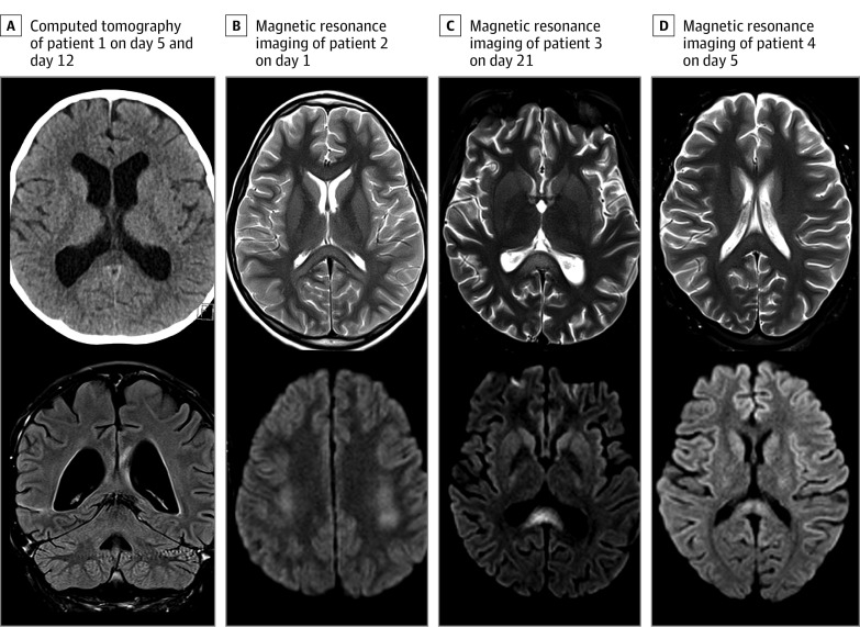
Results: Of the 27 children with COVID-19 pediatric multisystem inflammatory syndrome, 4 patients (14.8%) who were previously healthy had new-onset neurological symptoms. Symptoms included encephalopathy, headaches, brainstem and cerebellar signs, muscle weakness, and reduced reflexes. All 4 patients required intensive care unit admission for the treatment of COVID-19 pediatric multisystem inflammatory syndrome. Splenium signal changes were seen in all 4 patients on magnetic resonance imaging of the brain. In the 2 patients whose cerebrospinal fluid was tested, samples were acellular, with no evidence of infection on polymerase chain reaction or culture (including negative SARS-CoV-2 polymerase chain reaction results) and negative oligoclonal band test results. In all 3 patients who underwent electroencephalography, a mild excess of slow activity was found. Tests for N-methyl-d-aspartate receptor, myelin oligodendrocyte glycoprotein, and aquaporin-4 autoantibodies had negative results in all patients. In all 3 patients who underwent nerve conduction studies and electromyography, mild myopathic and neuropathic changes were seen. Neurological improvement was seen in all patients, with 2 making a complete recovery by the end of the study.
Conclusions and relevance: In this case-series study, children with COVID-19 presented with new neurological symptoms involving both the central and peripheral nervous systems and splenial changes on imaging, in the absence of respiratory symptoms. Additional research is needed to assess the association of neurological symptoms with immune-mediated changes among children with COVID-19.
Conflict of interest statement
Figures
References
MeSH terms
Supplementary concepts
Grants and funding
LinkOut - more resources
Full Text Sources
Other Literature Sources
Medical
Miscellaneous
