

doi: 10.1164/rccm.202004-1278IM.
Radiology-Pathology Correlation Demonstrating Organizing Pneumonia in a Patient Who Recovered from COVID-19
Affiliations
- PMID: 32609531
- PMCID: PMC7427386
- DOI: 10.1164/rccm.202004-1278IM
Item in Clipboard
Radiology-Pathology Correlation Demonstrating Organizing Pneumonia in a Patient Who Recovered from COVID-19
Am J Respir Crit Care Med.
.
No abstract available
Figures
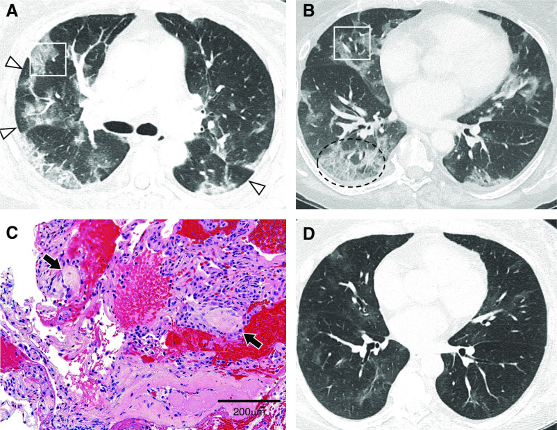
(A) Axial computed tomographic (CT) image of the midlung zones showing areas of “perilobular” distribution (arrowheads) of pulmonary opacities in a pattern consistent with organizing pneumonia. Approximate area of transbronchial biopsy in the anterior segment of the right upper lobe (white rectangle) is shown. (B) Axial CT image of the lower-lung zones showing patchy ground-glass opacities distributed peripherally in the lungs and “crazy paving” in the posterior basal right lower lobe (dashed oval). Approximate area of biopsy in the right middle lobe (white rectangle) is shown. (C) High-power magnification of alveolated lung containing scattered fibromyxoid plugs of granulation tissue (arrows) and sparse interstitial mononuclear infiltrates, indicating organizing pneumonia (hematoxylin and eosin). Scale bar, 200 μm. (D) Follow-up CT imaging performed 26 days after presentation with the axial image at the same location as in B, near the level of the inferior pulmonary veins, demonstrates nearly resolved pulmonary opacities. Videos 1 and 2 show the entirety of both CT scans.
References
LinkOut - more resources
Full Text Sources
