. 2020 Jul;24(7):1837-1857.
doi: 10.1109/JBHI.2020.2991043.

AI in Medical Imaging Informatics: Current Challenges and Future Directions

AI in Medical Imaging Informatics: Current Challenges and Future Directions

Andreas S Panayides et al. IEEE J Biomed Health Inform. 2020 Jul.

Abstract

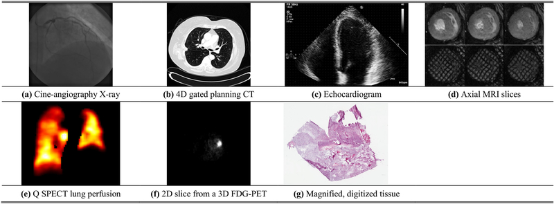
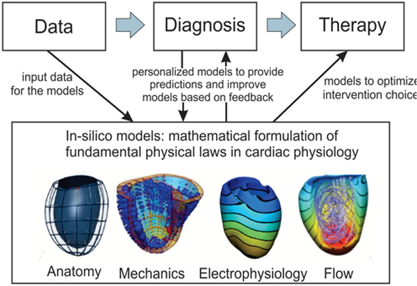
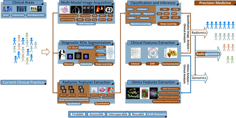
This paper reviews state-of-the-art research solutions across the spectrum of medical imaging informatics, discusses clinical translation, and provides future directions for advancing clinical practice. More specifically, it summarizes advances in medical imaging acquisition technologies for different modalities, highlighting the necessity for efficient medical data management strategies in the context of AI in big healthcare data analytics. It then provides a synopsis of contemporary and emerging algorithmic methods for disease classification and organ/ tissue segmentation, focusing on AI and deep learning architectures that have already become the de facto approach. The clinical benefits of in-silico modelling advances linked with evolving 3D reconstruction and visualization applications are further documented. Concluding, integrative analytics approaches driven by associate research branches highlighted in this study promise to revolutionize imaging informatics as known today across the healthcare continuum for both radiology and digital pathology applications. The latter, is projected to enable informed, more accurate diagnosis, timely prognosis, and effective treatment planning, underpinning precision medicine.


Figures

Fig. 1.
Fig. 1.
Typical medical imaging examples. (a) Cine angiography X-ray image after injection of iodinated contrast; (b) An axial slice of a 4D, gated planning CT image taken before radiation therapy for lung cancer; (c) Echocardiogram – 4 chamber view showing the 4 ventricular chambers (ventricular apex located at the top); (d) First row – axial MRI slices in diastole (left), mid-systole (middle), and peak systolic (right). Note the excellent contrast between blood pool and left ventricular myocardium. Second row –tissues tagged MRI slices at the same slice location and time point during the cardiac cycle. The modality creates noninvasive magnetic markers within the moving tissue [40]; (e) A typical Q SPECT image displaying lung perfusion in a lung-cancer patient; (f) A 2D slice from a 3D FDG-PET scan that shows a region of high glucose activity corresponding to a thoracic malignancy; (g) A magnified, digitized image of brain tissue to look for signs of Glioblastoma (taken from TCGA Glioblastoma Multiforme collection (https://cancergenome.nih.gov/).
Fig. 2.
Fig. 2.
In silico modelling paradigm of cardiovascular disease with application to heart.
Fig. 3.
Fig. 3.
Radiogenomics System Diagram: An abstract system diagram demonstrating the use of radiogenomics approaches in the context of precision medicine [68]. Based on the clinical case, (multi-modal) image acquisition is performed. Then, manual and/or automatic segmentation of the diagnostic regions of interest follows, driving quantitative and/or qualitative radiomic features extraction and machine learning approaches for segmentation, classification and inference. Alternatively, emerging deep learning methods using raw pixel intensities can be used for the same purpose. Radiogenomics approaches investigate the relationships between imaging and genomic features and how radiomics and genomics signatures, when processed jointly, can better describe clinical outcomes. On the other hand, radiomics research is focused on characterizing the relationship between quantitative imaging and clinical features.

References

    1. Kulikowski CA, “Medical imaging informatics: Challenges of definition and integration,” J. Amer. Med. Inform. Assoc, vol. 4, pp. 252–3, 1997. - PMC - PubMed
    1. Bui AA and Taira RK, Medical Imaging Informatics. Vienna, Austria: Springer, 2010.
    1. Society for Imaging Informatics in Medicine Web site, “Imaging informatics.” [Online]. Available: http://www.siimweb.org/index.cfm?id5324. Accessed: Jun. 2020.
    1. American Board of Imaging Informatics. [Online]. Available: https://www.abii.org/. Accessed: Jun. 2020.
    1. Hsu W, Markey MK, and Wang MD, “Biomedical imaging informatics in the era of precision medicine: Progress, challenges, and opportunities,” J. Amer. Med. Inform. Assoc, vol. 20, pp. 1010–1013, 2013. - PMC - PubMed

Publication types