. 2020 Jul 1;15(7):e0235546.
doi: 10.1371/journal.pone.0235546. eCollection 2020.

Systemic evaluation and localization of resistin expression in normal human tissues by a newly developed monoclonal antibody

Affiliations

Systemic evaluation and localization of resistin expression in normal human tissues by a newly developed monoclonal antibody

Qing Lin et al. PLoS One. .

Abstract

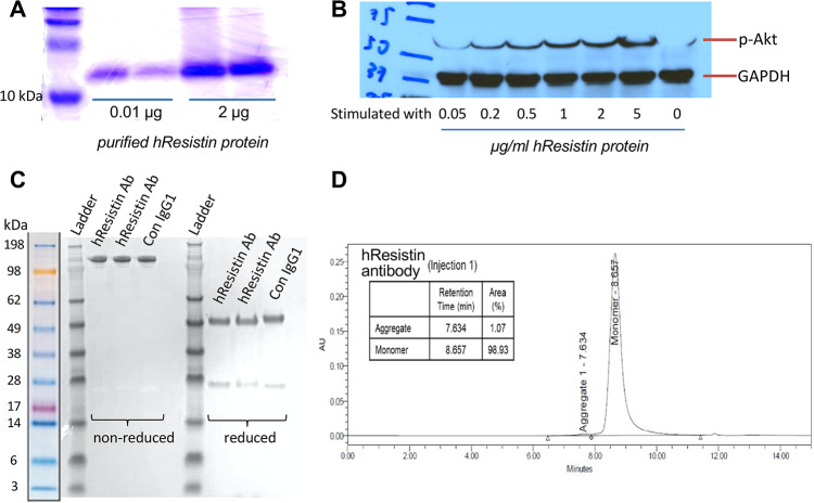
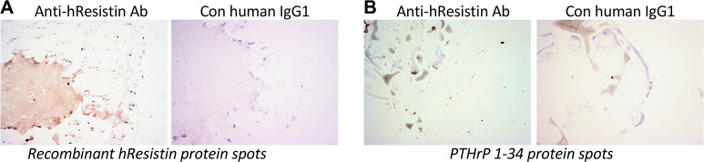
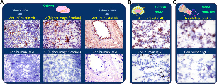
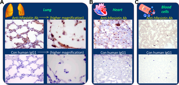
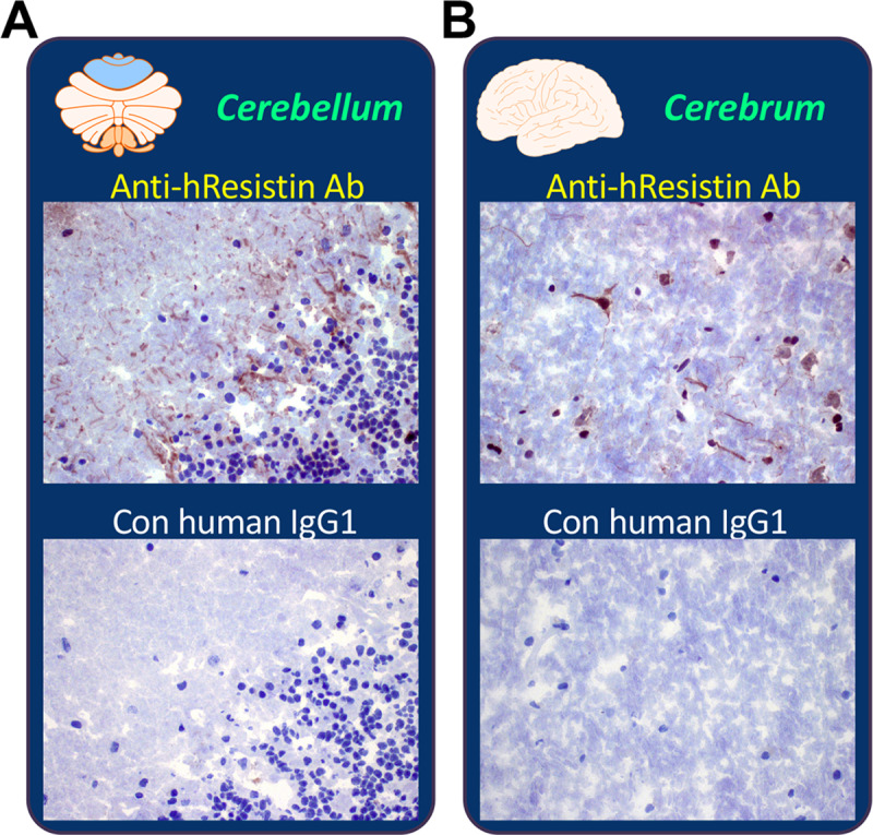
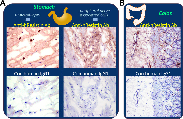
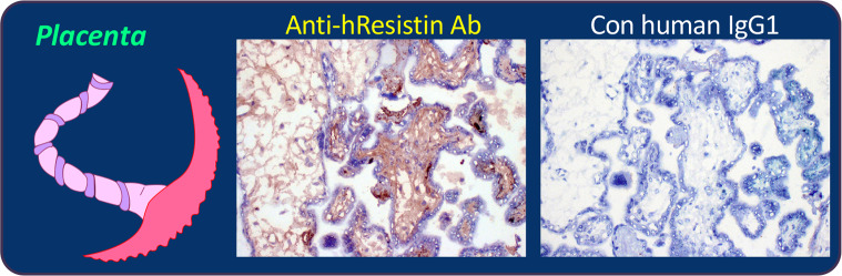
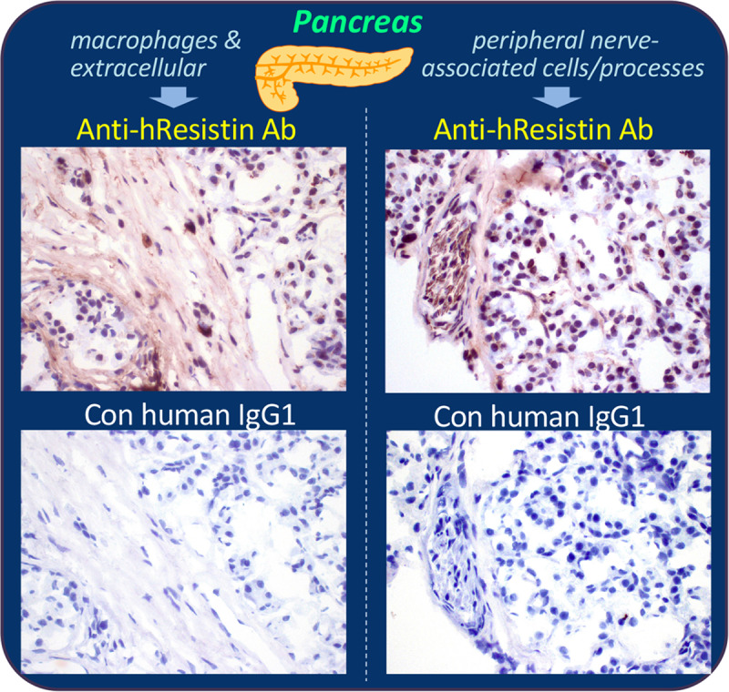
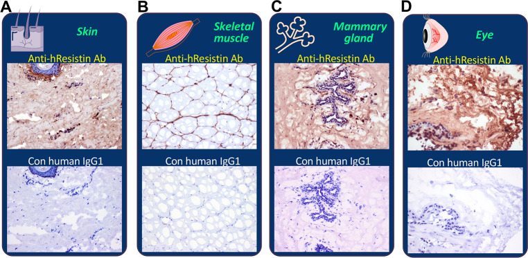
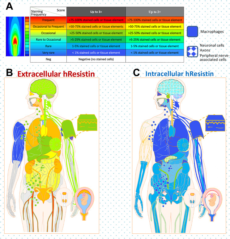
Resistin and resistin-like molecules are pleiotropic cytokines that are involved in inflammatory diseases. Our previous work suggested that resistin has the potential to be used as a biomarker and therapeutic target for human pulmonary arterial hypertension. However, data are limited on the distribution of resistin in healthy human organs. In this study, we used our newly developed anti-human resistin (hResistin) antibody to immunohistochemically detect the expression, localization, and intracellular/extracellular compartmentalization of hResistin in a full human tissue panel from healthy individuals. The potential cross reactivity of this monoclonal anti-hResistin IgG1 with normal human tissues also was verified. Results showed that hResistin is broadly distributed and principally localized in the cytoplasmic granules of macrophages scattered in the interstitium of most human tissues. Bone marrow hematopoietic precursor cells also exhibited hResistin signals in their cytoplasmic granules. Additionally, hResistin labeling was observed in the cytoplasm of nervous system cells. Notably, the cytokine activity of hResistin was illustrated by positively stained extracellular material in most human tissues. These data indicate that our generated antibody binds to the secreted hResistin and support its potential use for immunotherapy to reduce circulating hResistin levels in human disease. Our findings comprehensively document the basal expression patterns of hResistin protein in normal human tissues, suggest a critical role of this cytokine in normal and pathophysiologic inflammatory processes, and offer key insights for using our antibody in future pharmacokinetic studies and immunotherapeutic strategies.


Conflict of interest statement

The authors have read the journal’s policy and have the following potential competing interests: author SAP is a paid employee of Charles River Laboratories, Inc., a contract research organization that performed the immunopathological study to determine the tissue cross reactivity for the generated antihResistin antibody. The authors would like to declare the following patent applications associated with this research: RAJ had US (US 2016/0130341 A1) and international (WO 2014/204941 A1) patent applications pending for the monoclonal antibody developed against human resistin to cover pulmonary, cardiac, and other related inflammatory disorders. All other authors have declared that no competing interests exist. This does not alter our adherence to PLOS ONE policies on sharing data and materials.

Figures

Fig 1
Fig 1. Purity of the generated hResistin protein and anti-hResistin antibody.
(A) Coomassie-stained gel shows SDS-PAGE analysis of purified rhResistin protein. (B) Western blotting shows that the purified hResistin protein dose-dependently induced phosphorylation of Akt in the 3T3-L1 fibroblast cell line. (C) Purity of the anti-hResistin antibody and the control human IgG1 were analyzed by SDS-PAGE. (D) Size exclusion high-performance liquid chromatogram for the anti-hResistin antibody. Ab, antibody; Con, control; GAPDH, glyceraldehyde 3-phosphate dehydrogenase; hResistin, human resistin; p-Akt, phosphorylated Akt.
Fig 2
Fig 2. Representative images of immunohistochemically stained positive and negative control samples.
(A) Left: positive control rhResistin protein spots stained with 5 μg/mL anti-hResistin antibody (Ab). Weak to strong test antibody staining of proteinaceous material coincident with spotted positive control rhResistin is visible. Right: no staining was apparent when hResistin protein spots were stained with 5 μg/mL control (Con) human IgG1. (B) Left: Negative control PTHrP 1–34 protein spots stained with 5 μg/mL anti-hResistin antibody. No specific staining is apparent. Air bubbles and minor nonspecific staining at edge of dried material are noted. Right: PTHrP 1–34 protein spots stained with 5 μg/mL human IgG1. Control antibody staining is absent. Magnification: 100×.
Fig 3
Fig 3. Representative images of hResistin expression in the immune system.
(A) Human spleen tissues stained with 5 μg/mL anti-hResistin antibody. Left: anti-hResistin antibody stained cytoplasm/cytoplasmic granules in macrophages of germinal centers, whereas the same region exposed to 5 μg/mL control human IgG1 exhibited no staining. Right: anti-hResistin antibody stained the extracellular interstitium/stroma in perivascular areas, but control IgG1 did not stain any spleen tissue elements. Magnification: 400× (or higher: 600×). (B) Human lymph node tissues stained with 5 μg/mL anti-hResistin or control human antibodies. Magnification: 600×. (C) Human bone marrow tissues stained with 5 μg/mL anti-hResistin antibody or control IgG1. Magnification: 600×.
Fig 4
Fig 4. Representative images of hResistin expression in the cardiovascular-respiratory system.
Human lung tissue (A), heart tissue (B), and blood cells (C) stained with 5 μg/mL anti-hResistin or control human IgG1 antibodies. Blood cells were evaluated from peripheral blood smears. Antibody staining was also observed in the extracellular serum. Magnification: 400× (or higher: 600× in A).
Fig 5
Fig 5. Representative images of hResistin expression in the nervous system.
(A) Human brain cerebellum tissue stained with 5 μg/mL anti-hResistin antibody or control IgG1. Positive hResistin antibody staining was observed only rarely in the cytoplasm of axons. (B) Human brain cerebrum stained with 5 μg/mL anti-hResistin or control human IgG1. Cytoplasmic hResistin antibody staining of neuronal cell bodies and axons was rare. Magnification: 400×.
Fig 6
Fig 6. Representative images of hResistin expression in the digestive system.
(A) Human stomach tissue stained with 5 μg/mL anti-hResistin antibody. Left: h-Resistin antibody staining of cytoplasmic granules was observed in macrophages scattered in interstitium, whereas no staining was observed in the same region with 5 μg/mL control human IgG1. Right: h-Resistin antibody staining was observed in the cytoplasm of peripheral nerve-associated cells/processes in the myenteric plexus. Control IgG1 produced no such staining. Magnification: 400×. (B) Human colon tissue stained with 5 μg/mL anti-hResistin or control human IgG1. Magnification: 200×.
Fig 7
Fig 7. Representative images of hResistin expression in the urinary system.
Human kidney tissues were stained with 5 μg/mL anti-hResistin antibody (upper) or control IgG1 (lower). In the anti-hResistin antibody-stained tissue, extracellular positive signal was observed in interstitium/stroma perivascular areas. Anti-hResistin antibody did not stain vascular endothelium in the renal tissues. Control human IgG1 did not produce any staining in kidney tissue. Magnification: 200×.
Fig 8
Fig 8. Representative images of hResistin expression in the reproductive system.
Human placenta tissues stained with 5 μg/mL anti-hResistin antibody (left) or control IgG1 (right). Positive staining of secreted hResistin protein was observed in interstitium/stroma and aligned on extracellular matrix fibers. Control antibody did not produce any positive staining in human placenta. Magnification: 200×.
Fig 9
Fig 9. Representative images of hResistin expression in the endocrine system.
Human pancreas tissues stained with 5 μg/mL anti-hResistin antibody (upper panels) or control IgG1 (lower panels). Left: positive staining for hResistin was located in cytoplasmic granules of very rare macrophages scattered in interstitium, and in extracellular interstitium/stroma. Right: hResistin staining was also observed in cytoplasm of occasional cells/processes associated with peripheral nerves in some areas of pancreatic tissue. These regions did not exhibit staining by control human IgG1. Magnification: 400×.
Fig 10
Fig 10. Representative images of hResistin expression in the integumentary/muscular/sensory systems.
Tissues of human skin (A), striated skeletal muscle (B), mammary gland/breast (C), and eyes (D) were stained with 5 μg/mL anti-hResistin antibody or control human IgG1. Magnification: 200× (A, B, and D) or 400× (C).
Fig 11
Fig 11. Global analysis of hResistin expression in normal human organ tissues.
(A) Color coding (from cold [blue] to warm [red]) designates the positive intensity and frequency of anti-hResistin antibody staining. For the analysis of intracellular hResistin distribution, solid color-filled areas indicate the presence of hResistin-producing macrophages, whereas the pattern denotes hResistin-expressing peripheral nerve-associated cells, neuronal cells, or axons in the tissues. (B and C) Global mapping of the expression and distribution of extracellular (B) and intracellular (C) hResistin in normal human tissues.

Similar articles

Cited by

References

    1. Steppan CM, Bailey ST, Bhat S, Brown EJ, Banerjee RR, Wright CM, et al. The hormone resistin links obesity to diabetes. Nature. 2001;409(6818):307–12. 10.1038/35053000 . - DOI - PubMed
    1. Teng X, Li D, Champion HC, Johns RA. FIZZ1/RELMa, a novel hypoxia-induced mitogenic factor in lung with vasoconstrictive and angiogenic properties. Circ Res. 2003;92(10):1065–7. 10.1161/01.RES.0000073999.07698.33 . - DOI - PubMed
    1. Gerstmayer B, Kusters D, Gebel S, Muller T, Van Miert E, Hofmann K, et al. Identification of RELMg, a novel resistin-like molecule with a distinct expression pattern. Genomics. 2003;81(6):588–95. 10.1016/s0888-7543(03)00070-3 . - DOI - PubMed
    1. Holcomb IN, Kabakoff RC, Chan B, Baker TW, Gurney A, Henzel W, et al. FIZZ1, a novel cysteine-rich secreted protein associated with pulmonary inflammation, defines a new gene family. EMBO J. 2000;19(15):4046–55. 10.1093/emboj/19.15.4046 ; PMCID: PMC306596. - DOI - PMC - PubMed
    1. Steppan CM, Brown EJ, Wright CM, Bhat S, Banerjee RR, Dai CY, et al. A family of tissue-specific resistin-like molecules. Proc Natl Acad Sci U S A. 2001;98(2):502–6. 10.1073/pnas.98.2.502 ; PMCID: PMC14616. - DOI - PMC - PubMed

Publication types