. 2020 Nov:41:101048.
doi: 10.1016/j.molmet.2020.101048. Epub 2020 Jun 29.

Genetic loss of AMPK-glycogen binding destabilises AMPK and disrupts metabolism

Affiliations

Genetic loss of AMPK-glycogen binding destabilises AMPK and disrupts metabolism

Nolan J Hoffman et al. Mol Metab. 2020 Nov.

Abstract

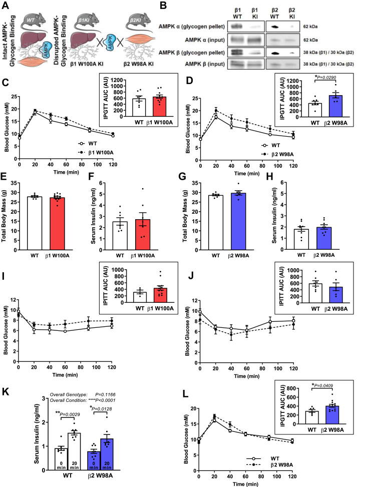
Objective: Glycogen is a major energy reserve in liver and skeletal muscle. The master metabolic regulator AMP-activated protein kinase (AMPK) associates with glycogen via its regulatory β subunit carbohydrate-binding module (CBM). However, the physiological role of AMPK-glycogen binding in energy homeostasis has not been investigated in vivo. This study aimed to determine the physiological consequences of disrupting AMPK-glycogen interactions.

Methods: Glycogen binding was disrupted in mice via whole-body knock-in (KI) mutation of either the AMPK β1 (W100A) or β2 (W98A) isoform CBM. Systematic whole-body, tissue and molecular phenotyping was performed in KI and respective wild-type (WT) mice.

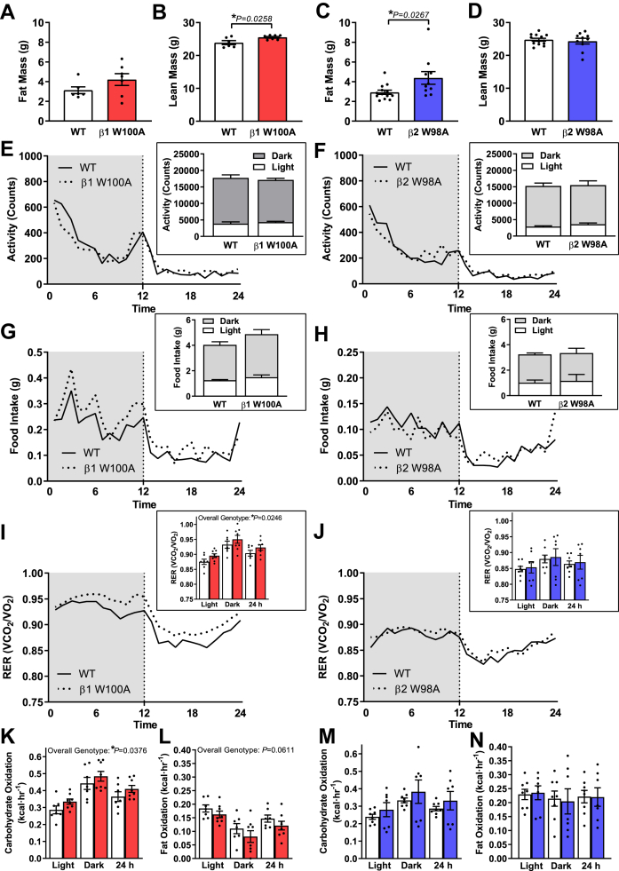
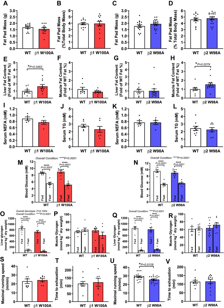
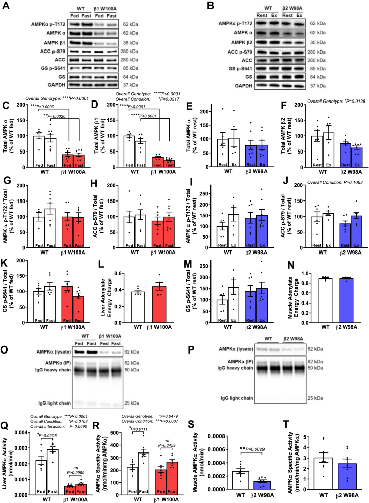
Results: While β1 W100A KI did not affect whole-body metabolism or exercise capacity, β2 W98A KI mice displayed increased adiposity and impairments in whole-body glucose handling and maximal exercise capacity relative to WT. These KI mutations resulted in reduced total AMPK protein and kinase activity in liver and skeletal muscle of β1 W100A and β2 W98A, respectively, versus WT mice. β1 W100A mice also displayed loss of fasting-induced liver AMPK total and α-specific kinase activation relative to WT. Destabilisation of AMPK was associated with increased fat deposition in β1 W100A liver and β2 W98A skeletal muscle versus WT.

Conclusions: These results demonstrate that glycogen binding plays critical roles in stabilising AMPK and maintaining cellular, tissue and whole-body energy homeostasis.

Keywords: AMP-activated protein kinase; Carbohydrate-binding module; Cellular energy sensing; Exercise; Liver; Skeletal muscle.


Figures

Figure 1
Figure 1
Disrupting AMPK β2 glycogen binding in vivo via KI mutation alters whole-body glucose handling. (A) Schematic depicts disruption of AMPK-glycogen binding in whole-body KI mice via mutation of the AMPK β1 subunit (W100A) and β2 subunit (W98A), predominantly expressed in liver and skeletal muscle, respectively. (B) Association of AMPK α (top panels) and AMPK β (bottom panels) from bacterially expressed AMPK containing β1 KI, β2 KI or respective WT with bovine liver glycogen relative to input protein (representative blots shown from 3 replicate experiments). Following 6-h fast, respective male WT and β1 W100A (C; n = 7–11) or β2 W98A (D; n = 6–7) age-matched mice (10–16 wk) were subjected to IPGTT (1 g/kg total body mass), and AUC was analysed (insets). (E) Total body mass (n = 7–11) and (F) serum insulin (n = 7) from WT and β1 W100A, or WT and β2 W98A (G) total body mass (n = 6–7) and (H) serum insulin (n = 8–9) was determined. Following 6-h fast, respective male WT and (I) β1 W100A (n = 4–12) or (J) β2 W98A (n = 6–7) age-matched mice (12–14 wk) were subjected to IPITT (0.5 units/kg total body mass) and AUC was analysed (insets). (K) Serum insulin was determined in male age-matched WT and β2 W98A mice (n = 7–8; 12–16 wk) at 0 min and 20 min following IP injection with 1 g/kg total body mass. Following a 6-h fast, male WT and β2 W98A age-matched mice (L; n = 6–12; 12–15 wk) were subjected to IPGTT (1 g/kg lean body mass), and AUC was analysed (inset). See also Figure S1. Data are represented as mean ± SEM; ∗P < 0.05; ∗∗P < 0.01; ∗∗∗∗P < 0.0001.
Figure 2
Figure 2
Disrupting AMPK β2 glycogen binding increases adiposity independent of changes in food intake, physical activity or patterns of whole-body fuel utilisation. Male age-matched mice (8–15 wk) were subjected to EchoMRI and CLAMS analyses. (A) Absolute fat and (b) absolute lean mass for WT and β1 W100A (n = 6–7) and WT and β2 W98A (C) absolute fat and (D) absolute lean mass (n = 10–13) are shown. (E) Infrared-based activity, (G) food intake and (I) RER (VO2/VCO2) were measured in WT and β1 W100A mice (n = 7–8). (F) Infrared-based activity, (H) food intake and (J) RER (VO2/VCO2) were measured in WT and β2 W98A mice (n = 7–8). Carbohydrate and fat oxidation were determined using RER from respective WT and β1 W100A (K and L; n = 7–8) or β2 W98A (M and N; n = 7–8) mice. See also Figure S2. Data are represented as mean ± SEM; ∗P < 0.05.
Figure 3
Figure 3
Loss of glycogen-bound AMPK β1 and β2 increases liver and skeletal muscle ectopic fat deposition independent of changes in fat pad mass, tissue glycogen or serum lipids. Tissues and serum were collected from male age-matched WT and KI mice (ranging from 14 to 32 wk). Epididymal fat pad (A) absolute mass and (B) mass relative to total body mass were determined in WT and β1 W100A mice (n = 12). Epididymal fat pad (C) absolute mass and (D) mass relative to total body mass were determined in WT and β2 W98A mice (n = 14–20). Freshly isolated (E) liver and (F) quadriceps muscle (n = 10 and n = 8–12, respectively) from WT and β1 W100A mice, and (G) liver and (H) muscle (n = 9–14 and n = 11–15, respectively) from WT and β2 W98A mice were subjected to EchoMRI. Following 6-h fast, colorimetric assays were used to determine serum (I) NEFA and (J) TG concentration (n = 7 and n = 5, respectively) in WT and β1 W100A and (K) NEFA and (L) TG concentration (n = 8–9 and n = 5–6, respectively) in WT and β2 W98A mice. (M) Blood glucose was measured, and glycogen content was determined using enzymatic analysis of (O) liver and (P) gastrocnemius muscle from fed or overnight fasted WT and W100A (n = 6–7). (N) Blood glucose and (Q) liver and (R) gastrocnemius muscle glycogen content are shown from fed or overnight fasted WT and β2 W98A mice (n = 6–10). Following treadmill acclimatisation, maximal running speed was determined in respective WT and (S) β1 W100A (n = 7–9) or (U) β2 W98A (n = 18) age-matched male mice (12–15 wk). Following 2–3 days' rest, time to exhaustion at 70% maximal speed was determined in respective WT and (T) β1 W100A (n = 6–9) or (V) β2 W98A (n = 11) mice. See also Figure S3. Data are represented as mean ± SEM; ∗P < 0.05; ∗∗∗∗P < 0.0001.
Figure 4
Figure 4
AMPK protein and kinase activity are reduced in liver and skeletal muscle of AMPK KI mice. Tissues were collected from male age-matched WT and KI mice (ranging from 12 to 22 wk; 14–32 wk for adenylate energy charge measurements). Representative immunoblots processed in parallel are shown for respective WT versus (A) fed and fasted β1 W100A liver or (B) gastrocnemius muscle from β2 W98A rested or treadmill exercised to exhaustion at 5° incline. Quantified immunoblots for total AMPK (C) α and (D) β from WT and β1 W100A liver (n = 4–7) and total AMPK (E) α and (F) β from WT and β2 W98A gastrocnemius muscle (n = 5–7) are shown. Quantified ratios of phosphorylated (G) AMPK T172, (H) ACC S79 and (K) GS S641 from fed and fasted WT and β1 W100A liver (n = 4–7), and phosphorylated (I) AMPK T172, (J) ACC S79 and (M) GS S641 from WT and β2 W98A gastrocnemius muscle (n = 5–7) relative to total protein are shown. Adenylate energy charge was determined via mass spectrometry analysis of AMP, ADP and ATP from respective WT and (L) β1 W100A liver (n = 5–6) or (N) β2 W98A gastrocnemius muscle (n = 5–6). Following AMPK α immunoprecipitation of respective WT and KI liver or muscle (O–P top panels, respectively; blots processed in parallel), total AMPK α activity was determined in (Q) fed and fasted β1 W100A liver (n = 6–7) and (S) rested β2 W98A gastrocnemius muscle via in vitro kinase assay using equal tissue input protein (n = 6–7). AMPK α-specific activity was determined in respective WT and (R) fed and fasted β1 W100A liver or (T) resting β2 W98A gastrocnemius muscle using balanced input of immunoprecipitated AMPK α determined from blots processed in parallel (O–P bottom panels; n = 6–7). See also Figure S4. Data are represented as mean ± SEM; ∗P < 0.05; ∗∗P < 0.01; ∗∗∗P < 0.001; ∗∗∗∗P < 0.0001.

Similar articles

Cited by

References

    1. Efeyan A., Comb W.C., Sabatini D.M. Nutrient-sensing mechanisms and pathways. Nature. 2015;517(7534):302–310. - PMC - PubMed
    1. Xiao B., Heath R., Saiu P., Leiper F.C., Leone P., Jing C. Structural basis for AMP binding to mammalian AMP-activated protein kinase. Nature. 2007;449(7161):496–500. - PubMed
    1. Scott J.W., Hawley S.A., Green K.A., Anis M., Stewart G., Scullion G.A. CBS domains form energy-sensing modules whose binding of adenosine ligands is disrupted by disease mutations. Journal of Clinical Investigation. 2004;113(2):274–284. - PMC - PubMed
    1. Townley R., Shapiro L. Crystal structures of the adenylate sensor from fission yeast AMP-activated protein kinase. Science. 2007;315(5819):1726–1729. - PubMed
    1. Polekhina G., Gupta A., Michell B.J., van Denderen B., Murthy S., Feil S.C. AMPK beta subunit targets metabolic stress sensing to glycogen. Current Biology. 2003;13(10):867–871. - PubMed

Publication types