. 2020 Sep;40(9):2279-2292.
doi: 10.1161/ATVBAHA.120.314491. Epub 2020 Jul 2.

Gut Microbiota Restricts NETosis in Acute Mesenteric Ischemia-Reperfusion Injury

Affiliations

Gut Microbiota Restricts NETosis in Acute Mesenteric Ischemia-Reperfusion Injury

Stefanie Ascher et al. Arterioscler Thromb Vasc Biol. 2020 Sep.

Abstract

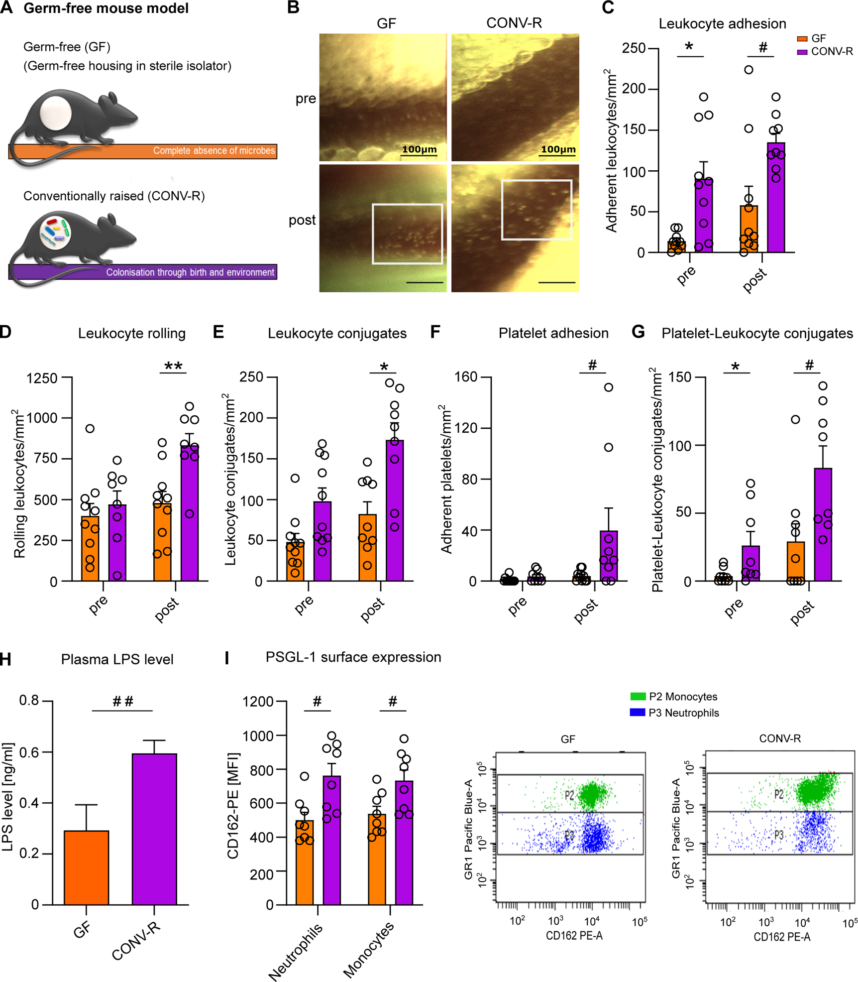
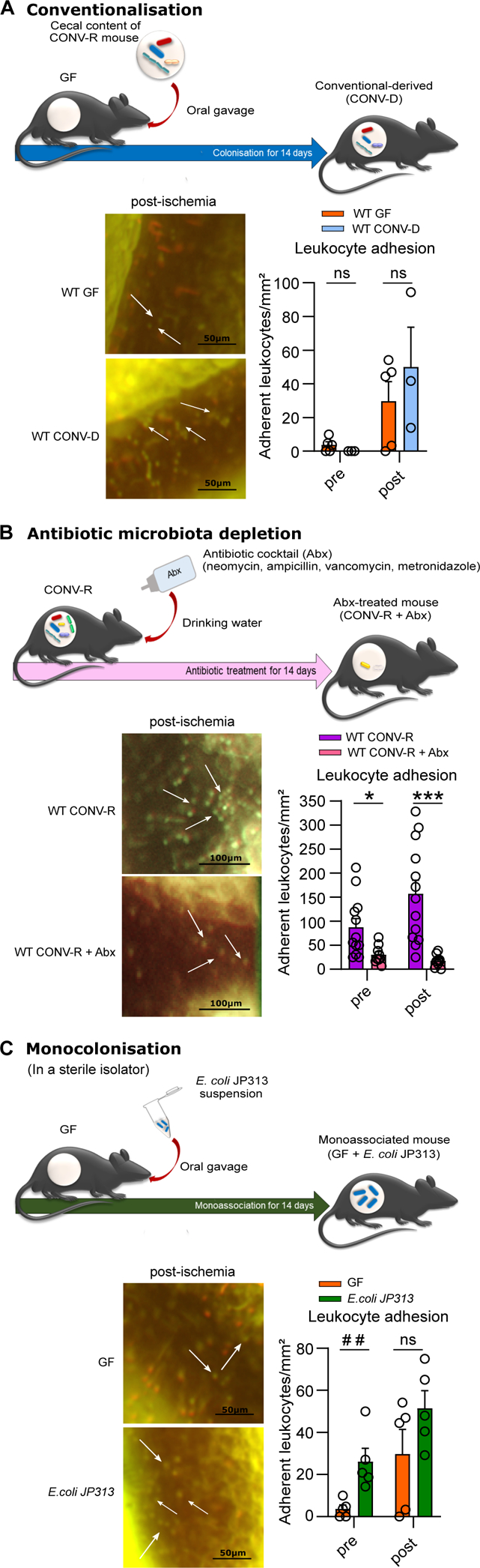
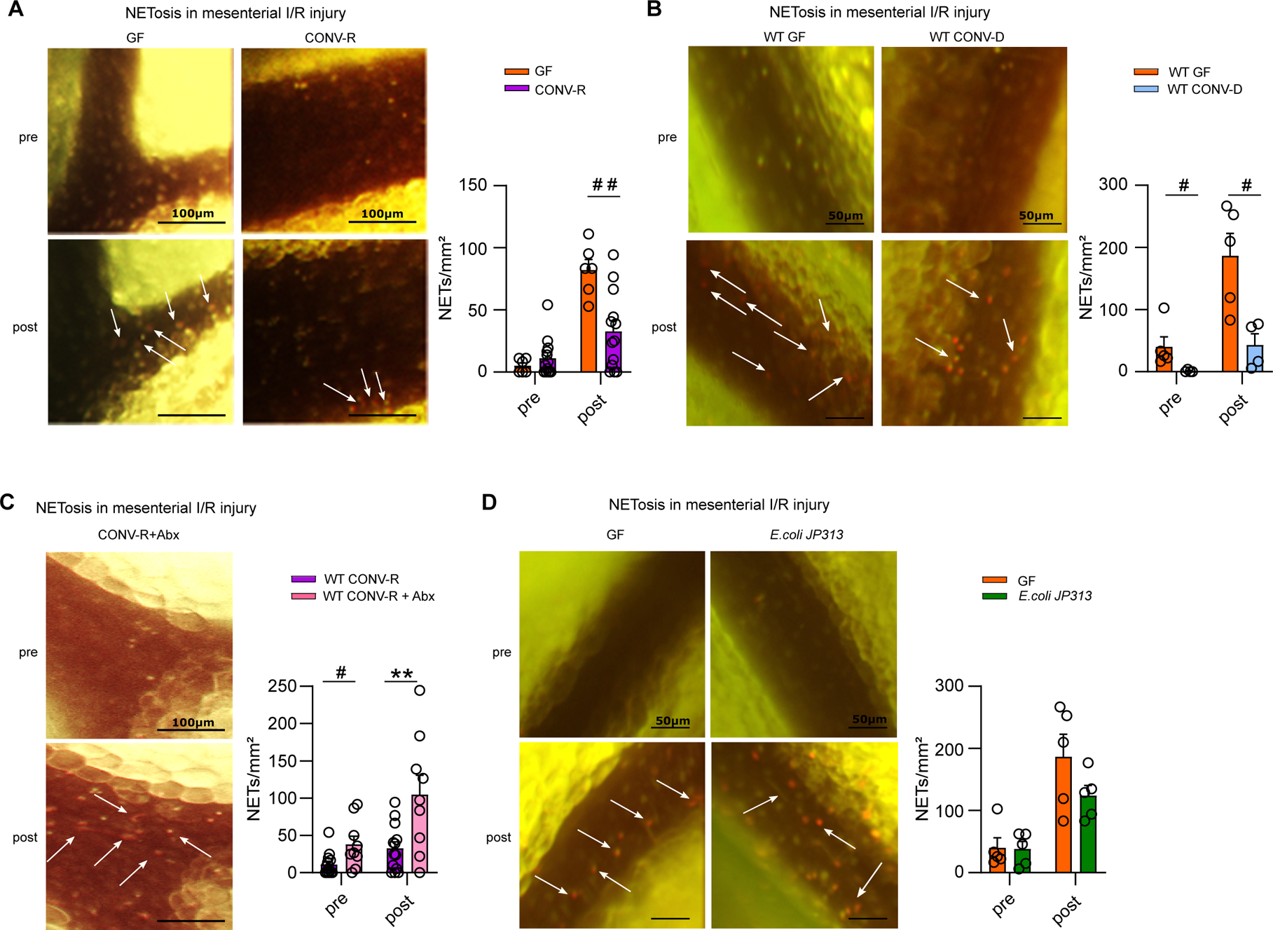
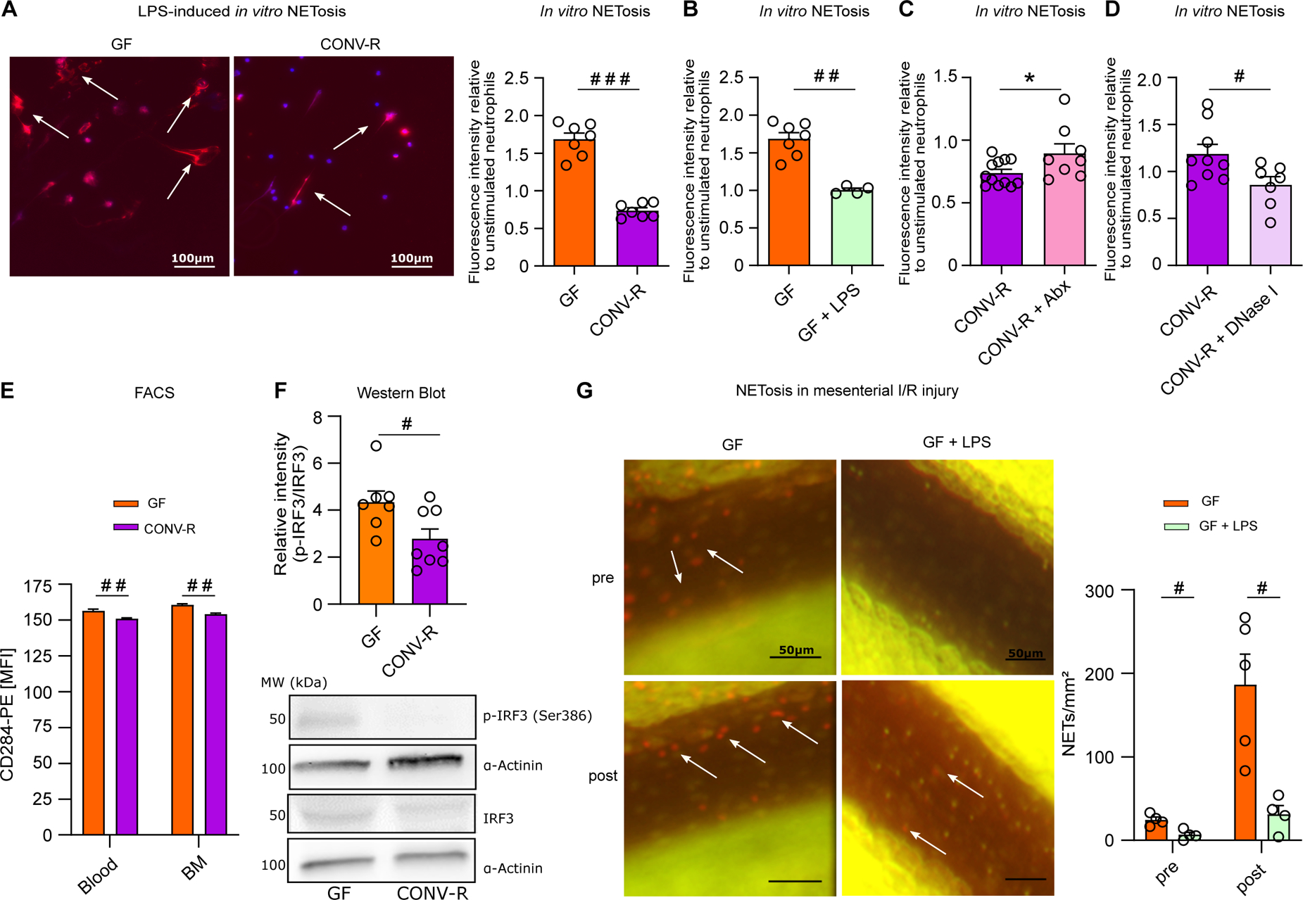
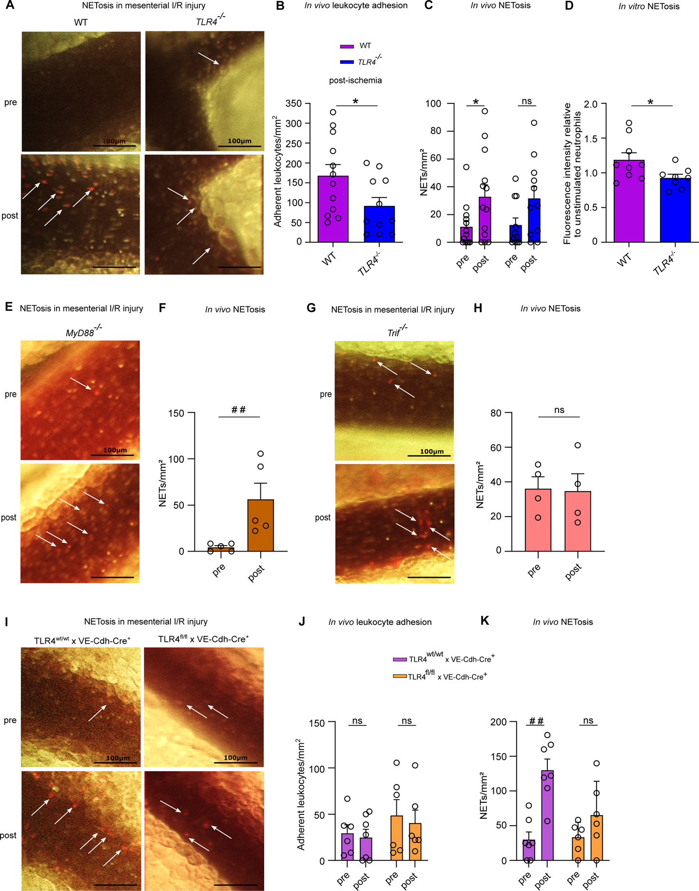
Objective: Recruitment of neutrophils and formation of neutrophil extracellular traps (NETs) contribute to lethality in acute mesenteric infarction. To study the impact of the gut microbiota in acute mesenteric infarction, we used gnotobiotic mouse models to investigate whether gut commensals prime the reactivity of neutrophils towards formation of neutrophil extracellular traps (NETosis). Approach and Results: We applied a mesenteric ischemia-reperfusion (I/R) injury model to germ-free (GF) and colonized C57BL/6J mice. By intravital imaging, we quantified leukocyte adherence and NET formation in I/R-injured mesenteric venules. Colonization with gut microbiota or monocolonization with Escherichia coli augmented the adhesion of leukocytes, which was dependent on the TLR4 (Toll-like receptor-4)/TRIF (TIR-domain-containing adapter-inducing interferon-β) pathway. Although neutrophil accumulation was decreased in I/R-injured venules of GF mice, NETosis following I/R injury was significantly enhanced compared with conventionally raised mice or mice colonized with the minimal microbial consortium altered Schaedler flora. Also ex vivo, neutrophils from GF and antibiotic-treated mice showed increased LPS (lipopolysaccharide)-induced NETosis. Enhanced TLR4 signaling in GF neutrophils was due to elevated TLR4 expression and augmented IRF3 (interferon regulatory factor-3) phosphorylation. Likewise, neutrophils from antibiotic-treated conventionally raised mice had increased NET formation before and after ischemia. Increased NETosis in I/R injury was abolished in conventionally raised mice deficient in the TLR adaptor TRIF. In support of the desensitizing influence of enteric LPS, treatment of GF mice with LPS via drinking water diminished LPS-induced NETosis in vitro and in the mesenteric I/R injury model.

Conclusions: Collectively, our results identified that the gut microbiota suppresses NETing neutrophil hyperreactivity in mesenteric I/R injury, while ensuring immunovigilance by enhancing neutrophil recruitment.

Keywords: extracellular traps; interferons; lipopolysaccharide; neutrophils; venules.


Conflict of interest statement

Disclosures

The authors declare no conflicts of interest.

Figures

Figure 1.
Figure 1.. Germ-free mice show reduced leukocyte adhesion to ischemia-reperfusion injured mesenteric venules.
(A) Germ-free (GF) and conventionally raised (CONV-R) mouse model (schematic representation). (B) Intravital imaging of mesenteric venules of GF and CONV-R mice pre- and post-ischemia; scale bar: 100 μm; representative images; white frame: acridine orange-stained adherent leukocytes. (C) Number of adhering leukocytes in mesenteric venules of GF and CONV-R mice (10 vs 9 mice/group) pre-ischemia and one hour post-ischemia. For GF and CONV-R male and female mice were used. (D) Number of rolling leukocytes pre-ischemia and one hour post-ischemia in GF and CONV-R mice (10 vs 8 mice/group). For GF and CONV-R male and female mice were used. (E) Number of leukocyte conjugates pre-ischemia and one hour post-ischemia in GF vs CONV-R mice (10 vs 9 mice/group). For GF and CONV-R male and female mice were used. (F) Number of adhering platelets pre-ischemia and one hour post-ischemia in GF and CONV-R mice (10 vs 9 mice/group). For GF and CONV-R male and female mice were used. (G) Number of platelet-leukocyte conjugates pre-ischemia and one hour post-ischemia in GF and CONV-R (9 vs 8 mice/group). For GF and CONV-R male and female mice were used. (G) Plasma LPS levels in GF vs CONV-R mice (6 vs 10 mice/group). For GF and CONV-R male and female mice were used. (H, I) FACS analysis on PSGL-1 surface expression on monocytes (green) and neutrophils (blue) in whole blood of GF and CONV-R mice (8 vs 8 mice/group). For GF and CONV-R male and female mice were used. Results are shown as means ± s.e.m. Scale bar: 100 μm. Statistical comparisons were performed using the independent samples Studentś t-test (*) or the Mann-Whitney test (#), */#p<0.05, **/##p<0.01.
Figure 2.
Figure 2.. Colonization with a gut microbiota or monocolonization with Escherichia coli augments leukocyte adhesion to the ischemia-reperfusion injured endothelium of mesenteric venules.
(A) Number of adhering leukocytes in mesenteric venules of germ-free (GF) and conventional-derived (CONV-D) mice (5 vs 4 mice/group) pre-ischemia and one hour post-ischemia; adhering leukocytes were stained with acridine orange. For WT GF male mice and WT CONV-D male and female mice were used. (B) Number of adhering leukocytes pre-ischemia and post-ischemia in conventionally raised (CONV-R) controls and CONV-R antibiotic (Abx)-treated mice (13 vs 9 mice/group); adhering leukocytes were stained with acridine orange. For WT CONV-R and CONV-R (Abx)-treated male and female mice were used. (C) Number of adhering leukocytes pre-ischemia and post-ischemia in GF and GF monocolonized with E. coli JP313 mice (5 vs 5 mice/group); adhering leukocytes were stained with acridine orange. For GF male mice and for GF monocolonized with E. coli JP313 male and female mice were used. Results are shown as means ± s.e.m. Scale bar: 50 μm or 100 μm. Statistical comparisons were performed using the independent samples Studentś t-test (*) or the Mann-Whitney test (#), */#p<0.05, **/##p<0.01, ***/### p<0.001.
Figure 3.
Figure 3.. The presence of gut commensals restricts the formation of neutrophil extracellular traps (NETosis) in ischemia-reperfusion injured mesenteric venules.
(A) NETosis in mesenteric venules of GF and CONV-R mice (6 vs 13 mice/group) pre- and post-ischemia; adhering leukocytes were stained with acridine orange; NETs were visualized by SYTOX orange. For GF and CONV-R male and female mice were used. (B) NETosis in mesenteric venules pre- and post-ischemia in GF and CONV-D mice (5 vs 4 mice/group); adhering leukocytes were stained with acridine orange; NETs were visualized by SYTOX orange. For WT GF male mice and WT CONV-D male and female mice were used. (C) NETosis in mesenteric venules pre- and post-ischemia in CONV-R and (Abx)-treated CONV-R mice (13 vs 9 mice/group); adhering leukocytes were stained with acridine orange; NETs were visualized by SYTOX orange. For WT CONV-R and CONV-R (Abx)-treated male and female mice were used. (D) NETosis in mesenteric venules pre- and post-ischemia in GF and GF mice monoassociated with E.coli JP 313 (5 vs 5 mice/group); adhering leukocytes were stained with acridine orange; NETs were visualized by SYTOX orange. For GF male mice and GF monoassociated with E.coli JP 313 male and female mice were used. Results are shown as means ± s.e.m. Scale bar: 50 μm or 100 μm. Statistical comparisons were performed using the independent samples Studentś t-test (*) or the Mann-Whitney test (#), */#p<0.05, **/##p<0.01.
Figure 4.
Figure 4.. Hyperreactive TLR4 signaling promotes enhanced NETosis in the germ-free mouse model.
(A) Representative images of LPS-induced in vitro NET formation; nuclear staining (DAPI; blue); NETs (visualized by citrullinated histone H3 antibody staining; red); scale bar: 100 μm. LPS-induced in vitro NETosis of cultured bone marrow neutrophils from GF and CONV-R mice (7 vs 7 mice/group). For GF and CONV-R male and female mice were used. (B) LPS-induced in vitro NETosis of cultured bone marrow neutrophils from GF and LPS-treated GF mice (7 vs 4 mice/group). For GF and LPS-treated GF male and female mice were used. (C) LPS-induced in vitro NETosis of cultured bone marrow neutrophils from CONV-R and (Abx)-treated CONV-R mice (12 vs 8 mice/group). For CONV-R and (Abx)-treated CONV-R male and female mice were used. (D) LPS-induced in vitro NETosis of cultured bone marrow neutrophils from CONV-R and DNase I-treated CONV-R control mice (9 vs 7 mice/group). For CONV-R and DNase I-treated CONV-R controls male and female mice were used. (E) FACS analysis on CD284 (TLR4) surface expression on neutrophils in whole blood and bone marrow of GF and CONV-R mice. For GF and CONV-R male and female mice were used. (F) Western Blot analysis of relative intensity of phosphorylated IRF-3 level to total IRF-3 protein level in GF and CONV-R mice; results are normalized to α-actinin. For GF and CONV-R male and female mice were used. (G) NETosis in mesenteric venules pre- and post-ischemia in GF and LPS-treated GF mice (5 vs 4 mice/group). For GF male mice and LPS-treated GF male and female mice were used. Results are shown as means ± s.e.m. Scale bar: 50 μm or 100 μm. Statistical comparisons were performed using the independent samples Studentś t-test (*) or the Mann-Whitney test (#), */#p<0.05, **/##p<0.01, ***/###p<0.001.
Figure 5.
Figure 5.. The TLR4/TRIF signaling axis is critically involved in mesenteric I/R injury-induced NETosis.
(A) Intravital imaging of mesenteric venules of CONV-R WT and CONV-R TLR4-deficient mice pre- and post-ischemia; scale bar: 100 μm; adhering leukocytes were stained with acridine orange; NETs were visualized by SYTOX orange. (B) Number of adhering leukocytes one hour post-ischemia in CONV-R WT and CONV-R TLR4-deficient mice (12 vs 12 mice/group); adhering leukocytes were stained with acridine orange. For CONV-R WT and CONV-R TLR4−/− male and female mice were used. (C) NETosis in mesenteric venules pre- and post-ischemia in CONV-R WT and CONV-R TLR4-deficient mice (13 vs 11 mice/group). For CONV-R WT and CONV-R TLR4−/− male and female mice were used. (D) LPS-induced in vitro NETosis of cultured bone marrow neutrophils from CONV-R WT and CONV-R TLR4-deficient mice (9 vs 8 mice/group). For CONV-R WT and CONC-R TLR4−/− male and female mice were used. (E) Intravital imaging of mesenteric venules of CONV-R MyD88-deficient mice pre- and post-ischemia; scale bar: 100 μm; adhering leukocytes were stained with acridine orange; NETs were visualized by SYTOX orange. (F) NETosis in mesenteric venules pre- and post-ischemia in MyD88-deficient mice (5 mice; male and female mice were used). (G) Intravital imaging of mesenteric venules of Trif-deficient mice pre- and post-ischemia; scale bar: 100 μm; adhering leukocytes were stained with acridine orange; NETs were visualized by SYTOX orange. (H) NETosis in mesenteric venules pre- and post-ischemia in Trif-deficient mice (4 mice; male and female mice were used). (I) Intravital imaging of mesenteric venules of TLR4-flox × VE-Cadherin-Cre (deficient in endothelial TLR4) mice and their controls pre- and post-ischemia; scale bar: 100 μm; adhering leukocytes were stained with acridine orange; NETs were visualized by SYTOX orange. (J) Number of adhering leukocytes pre-ischemia and one hour post-ischemia in TLR4-flox × VE-Cadherin-Cre (deficient in endothelial TLR4) mice and their controls (7 vs 6 mice/group). For TLR4-flox × VE-Cadherin-Cre Controls female and for TLR4-flox × VE-Cadherin-Cre female and male mice were used. (K) NETosis in mesenteric venules pre- and post-ischemia in TLR4-flox × VE-Cadherin-Cre (deficient in endothelial TLR4) mice and their controls (7 vs 6 mice/group). For TLR4-flox × VE-Cadherin-Cre Controls female and for TLR4-flox × VE-Cadherin-Cre female and male mice were used. Results are shown as means ± s.e.m. Scale bar: 100 μm. Statistical comparisons were performed using the independent samples Studentś t-test (*) or the Mann-Whitney test (#), */#p<0.05, **/##p<0.01.

Comment in

References

    1. Walker TG. Mesenteric ischemia. Semin Intervent Radiol. 2009; 26:175–183. doi: 10.1055/s-0029-1225662 - DOI - PMC - PubMed
    1. Harward TR., Brooks DL, Flynn TC, Seeger JM. Multiple organ dysfunction after mesenteric artery revascularization. J Vasc Surg. 1993; 18:459–467. doi: 10.1067/mva.1993.48586 - DOI - PubMed
    1. Shigematsu T, Wolf RE, Granger DN. T-lymphocytes modulate the microvascular and inflammatory responses to intestinal ischemia-reperfusion. Microcirculation 2002; 9:99–109. doi: 10.1038/sj/mn/7800126 - DOI - PubMed
    1. Schoenberg MH, Berger HG. Reperfusion injury after intestinal ischemia. Crit Care Med. 1993; 21:1376–1386. doi: 10.1097/00003246-199309000-00023 - DOI - PubMed
    1. Cicalese L, Billiar TR, Rao AS, Bauer AJ. Interaction between ischemia/reperfusion-induced leukocyte emigration and translocating bacterial enterotoxins on enteric muscle function. Transplant Proc. 1997; 29:1815. doi: 10.1016/s0041-1345(97)00080-8 - DOI - PubMed

Publication types

MeSH terms