. 2020 Jul 1;20(1):202.
doi: 10.1186/s12906-020-02961-0.

Phyllanthus amarus prevents LPS-mediated BV2 microglial activation via MyD88 and NF-κB signaling pathways

Affiliations

Phyllanthus amarus prevents LPS-mediated BV2 microglial activation via MyD88 and NF-κB signaling pathways

Elysha Nur Ismail et al. BMC Complement Med Ther. .

Abstract

Background: Phyllanthus amarus has been shown to attenuate lipopolysaccharide (LPS)-induced peripheral inflammation but similar studies in the central nervous system are scarce. The aim of the present study was to investigate the neuroprotective effects of 80% ethanol extract of P. amarus (EPA) in LPS-activated BV2 microglial cells.

Methods: BV2 microglial cells c for 24 h, pre-treated with EPA for 24 h prior to LPS induction for another 24 h. Surface expression of CD11b and CD40 on BV2 cells was analyzed by flow cytometry. ELISA was employed to measure the production of pro-inflammatory mediators i.e. nitric oxide (NO) and tumor necrosis factor (TNF)-α. Western blotting technique was used to determine the expression of inducible nitric oxide synthase (iNOS), myeloid differentiation protein 88 (MYD88), nuclear factor kappa B (NF-κB), caspase-1, and mitogen activated protein kinase (MAPK).

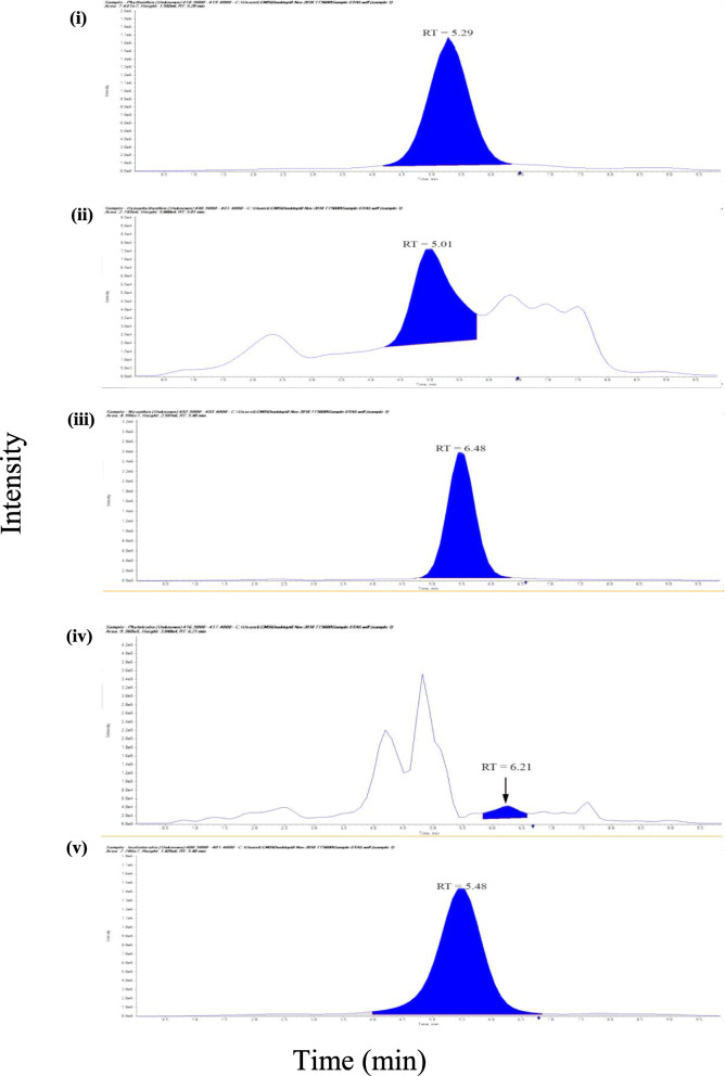
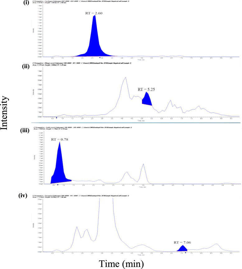
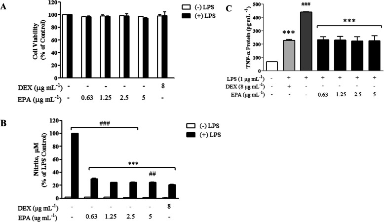
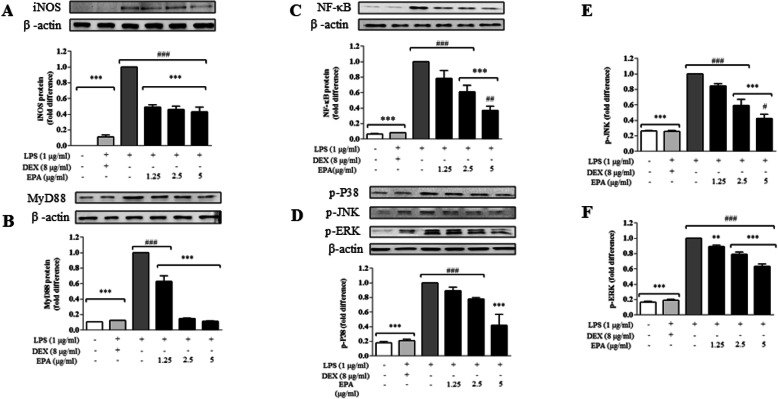
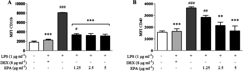
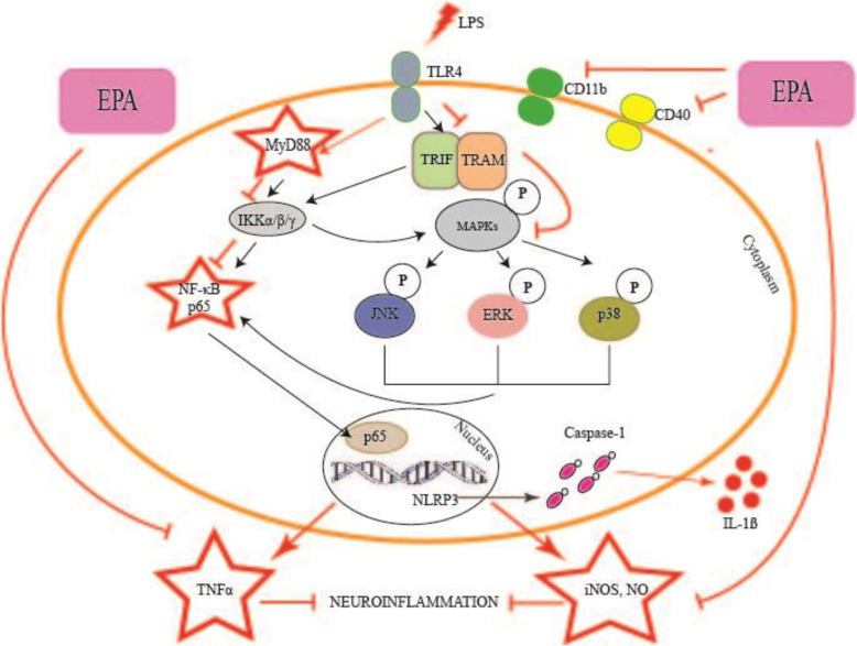
Results: Qualitative and quantitative analyses of the EPA using a validated ultra-high pressure liquid chromatography tandem mass spectrometry (UHPLC-MS/MS) method indicated the presence of phyllanthin, hypophyllanthin, niranthin, ellagic acid, corilagin, gallic acid, phyltetralin, isolintetralin and geraniin. EPA suppressed the production of NO and TNFα in LPS-activated BV2 microglial cells. Moreover, EPA attenuated the expression of MyD88, NF-κB and MAPK (p-P38, p-JNK and p-ERK1/2). It also inhibited the expression of CD11b and CD40. EPA protected against LPS-induced microglial activation via MyD88 and NF-κB signaling in BV2 microglial cells.

Conclusions: EPA demonstrated neuroprotective effects against LPS-induced microglial cells activation through the inhibition of TNFα secretion, iNOS protein expression and subsequent NO production, inhibition of NF-κB and MAPKs mediated by adapter protein MyD88 and inhibition of microglial activation markers CD11b and CD40.

Keywords: BV2 microglial cells; Microglial activation; Neuroinflammation; Neuroprotection; Phyllanthus amarus.


Conflict of interest statement

The authors declare that they have no competing interests.

Figures

Fig. 1
Fig. 1
Positive mode: MRM chromatogram of reference standard, lignans. (i) phyllanthin, (ii) hypophyllanthin, (iii) niranthin, (iv) phyltetralin and (v) isolintetralin. RT: retention time
Fig. 2
Fig. 2
Negative mode MRM chromatogram of reference standard, hydrolysable tannins. (i) corilagin, (ii) ellagic acid, (iii) gallic acid and (iv) geraniin. RT: retention time
Fig. 3
Fig. 3
Effects of EPA on cell viability of BV2 cells. Cells were treated with the extract at indicated concentrations or 1 μg ml-1 LPS for 24 h. a Cell viability was examined with MTT assay, b Nitric oxide analysis with Griess Assay and (c) TNFα analysis using ELISA. Results were expressed as the percentage of surviving cells relative to control cells and presented as mean ± SEM of three independent experiments; each was performed in triplicate. Cell viability of treated cells was compared to the respective control sample (cell treated with same amount of vehicle). Statistical differences between groups were assessed by one-way analysis of variance (ANOVA) from at least three independent experiments, followed by Dunnett’s test.* indicate means significantly differ at p < 0.05
Fig. 4
Fig. 4
EPA attenuated iNOS via MyD88/MAP kinase pathway, cells were induced with LPS (1 μg ml− 1) for 24 h in the presence of EPA. Western blots detecting iNOS (a), MyD88 (b) NFκB (c), (d) p-P38, (e) p-JNK and (f) p-ERK was performed as described in the methodology. Data are shown as mean ± SEM. Statistical differences between groups were assessed by one-way analysis of variance (ANOVA) from at least three independent experiments, followed by Dunnett’s test. * indicates p < 0.05 as compared to LPS and # indicates p < 0.05 as compared to DEX
Fig. 5
Fig. 5
Expression of CD11b (a) and CD40 (b) on BV2 cells after 24 h LPS-induction prior to a 24 h treatment with EPA or DEX. Statistical analysis of median fluorescence intensity (MFI) of the markers shown in A and B are expressed as mean ± SEM. Statistical differences between groups were assessed by one-way analysis of variance (ANOVA) from at least three independent experiments, followed by Dunnett’s test. * indicates p < 0.05 as compared to LPS and # indicates p < 0.05 as compared to DEX
Fig. 6
Fig. 6
The proposed neuroprotective effects of EPA in BV2 microglial cells challenged with LPS through inhibition of the processes involved in neuroinflammation. EPA: ethanolic P. amarus extract, LPS: lipopolysaccharide, TLR4: toll-like receptor 4, TRAF6: THF receptor associated factor 6, IKK: IκB kinase, MAPK: mitogen activated protein kinase, JNK: c-Jun N-terminal kinase, ERK: extracellular-signal-regulated kinase, P38: p38 mitogen-activated protein kinase, NF-κB:nuclear factor kappa light chain enhancer of activated B cells, TNFα: tumor necrosis factor alpha, IL: interleukin (e.g., IL-1β, IL-6), iNOS: inducible nitric oxide synthase, PGE2: prostaglandin E2, COX-2: cyclooxygenase-2

References

    1. Wang X, Hu D, Zhang L, Lian G, Zhao S, Wang C, et al. Gomisin A inhibits lipopolysaccharide-induced inflammatory responses in N9 microglia via blocking the NF-κB/MAPKs pathway. Food Chem Toxicol. 2014;63:119–127. - PubMed
    1. Kofler J, Wiley CA. Microglia. Toxicol Pathol. 2011;39(1):103–114. - PubMed
    1. Labzin LI, Heneka MT, Latz E. Innate Immunity and Neurodegeneration. Annu Rev Med. 2018;69(1):437–449. - PubMed
    1. Kettenmann H, Hanisch U, Noda M, Verkhratsky A. Physiology of Microglia. Physiol Rev. 2011;91:461–553. - PubMed
    1. Amor S, Peferoen LAN, Vogel DYS, Breur M, van der Valk P, Baker D, et al. Inflammation in neurodegenerative diseases - an update. Immunology. 2014;142(2):151–166. - PMC - PubMed