

A 3D-printed, personalized, biomechanics-specific beta-tricalcium phosphate bioceramic rod system: personalized treatment strategy for patients with femoral shaft non-union based on finite element analysis
- PMID: 32611412
- PMCID: PMC7331224
- DOI: 10.1186/s12891-020-03465-1
A 3D-printed, personalized, biomechanics-specific beta-tricalcium phosphate bioceramic rod system: personalized treatment strategy for patients with femoral shaft non-union based on finite element analysis
Abstract
Background: Although double-plate fixation (DP), i.e., fixation with a combination of a main lateral plate (LP) and a support medial plate (MP), is a relatively mature method for treating femoral shaft non-union with bone defect causes complications. The purpose of this study was to evaluate LP fixation with a 3D-printed, personalized, biomechanics-specific β-TCP bioceramic rod system (LP + 3DpbsBRS) as an alternative with less collateral damage.
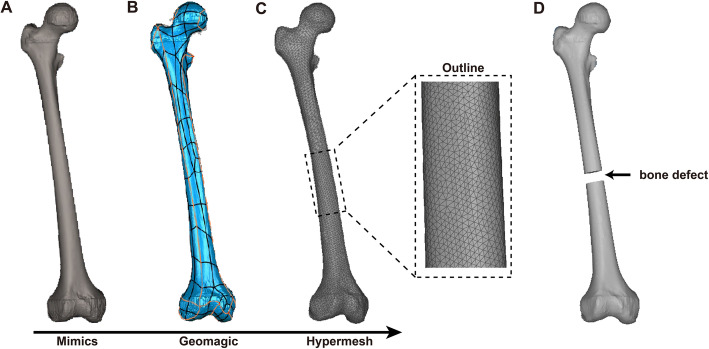
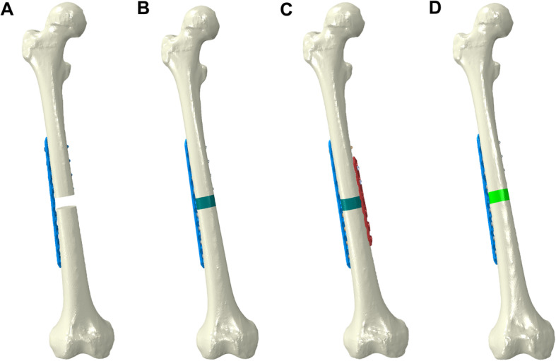
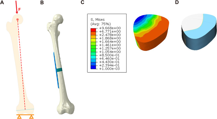
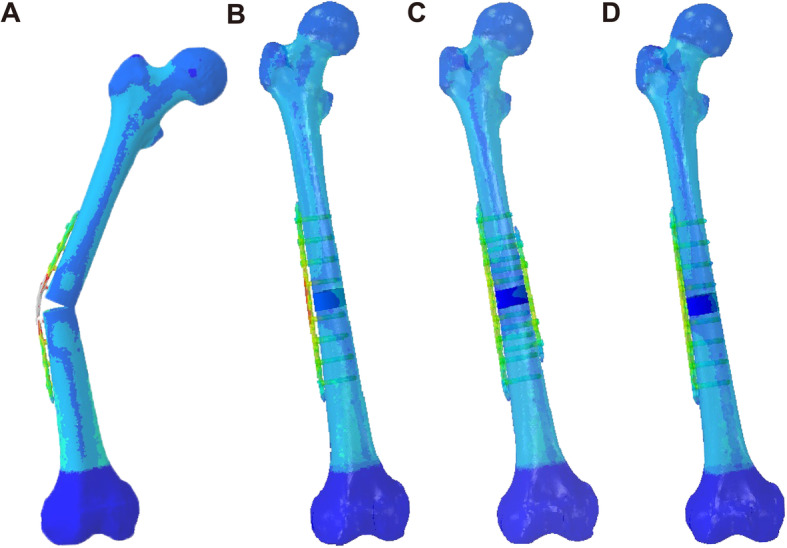
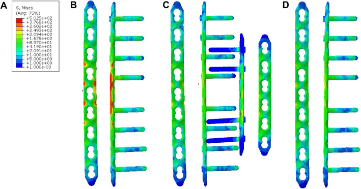
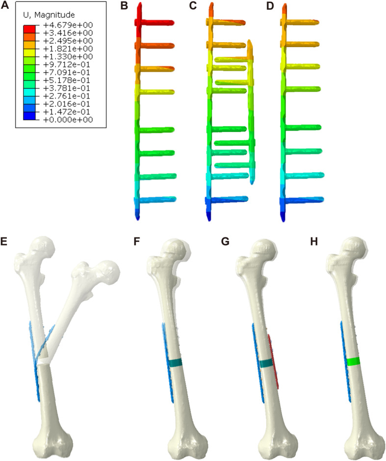
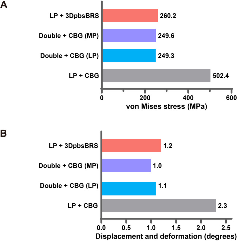
Methods: Structure-specific finite element modelling was used to simulate femoral shaft non-union with bone defects and treatment with an LP only as the blank control. Then, the peak von Mises stress (VMS), the VMS distribution, and the plate displacement were determined to compare the effectiveness of LP + CBG (cancellous bone grafting), DP + CBG, and LP + 3DpbsBRS under 850 N of axial force.
Results: Our results indicated that the peak VMS was 260.2 MPa (LP + 3DpbsBRS), 249.6 MPa (MP in DP + CBG), 249.3 MPa (LP in DP + CBG), and 502.4 MPa (LP + CBG). The bending angle of the plate was 1.2° versus 1.0° versus 1.1° versus 2.3° (LP + 3DpbsBRS versus MP in DP + CBG versus LP in DP + CBG versus LP + CBG).
Conclusion: The 3DpbsBRS in the LP + 3DpbsBRS group could replace the MP in the DP + CBG group by providing similar medial mechanical support. Furthermore, avoiding the use of an MP provides better protection of the soft tissue and vasculature.
Conflict of interest statement
The authors declare that they have no competing interests.
Figures
Similar articles
-
Plate configuration for biological reconstructions of femoral intercalary defect - a finite element evaluation.Comput Methods Programs Biomed. 2022 Sep;224:107006. doi: 10.1016/j.cmpb.2022.107006. Epub 2022 Jul 4. Comput Methods Programs Biomed. 2022. PMID: 35816816
-
Finite element analysis of different medial fixation strategies in double-plate osteosynthesis for AO type 33-C2 Femoral fractures.Injury. 2023 Apr;54 Suppl 2:S86-S94. doi: 10.1016/j.injury.2022.01.003. Epub 2022 Jan 5. Injury. 2023. PMID: 35219538
-
Biomechanical assessment of single LISS versus double-plate osteosynthesis in the AO type 33-C2 fractures: A finite element analysis.Injury. 2018 Dec;49(12):2142-2146. doi: 10.1016/j.injury.2018.10.011. Epub 2018 Oct 9. Injury. 2018. PMID: 30322705
-
Finite Element Analysis of Different Double-Plate Angles in the Treatment of the Femoral Shaft Nonunion with No Cortical Support opposite the Primary Lateral Plate.Biomed Res Int. 2018 Jul 31;2018:3267107. doi: 10.1155/2018/3267107. eCollection 2018. Biomed Res Int. 2018. PMID: 30151378 Free PMC article.
-
Biomechanical and biological aspects of defect treatment in fractures using helical plates.Acta Chir Orthop Traumatol Cech. 2014;81(4):267-71. Acta Chir Orthop Traumatol Cech. 2014. PMID: 25137496 Review.
Cited by
-
Beyond hype: unveiling the Real challenges in clinical translation of 3D printed bone scaffolds and the fresh prospects of bioprinted organoids.J Nanobiotechnology. 2024 Aug 21;22(1):500. doi: 10.1186/s12951-024-02759-z. J Nanobiotechnology. 2024. PMID: 39169401 Free PMC article. Review.
-
Co-modified 3D printed β-tricalcium phosphate with magnesium and selenium promotes bone defect regeneration in ovariectomized rat.J Mater Sci Mater Med. 2023 Jan 9;34(1):7. doi: 10.1007/s10856-022-06708-w. J Mater Sci Mater Med. 2023. PMID: 36622473 Free PMC article.
References
MeSH terms
Substances
LinkOut - more resources
Full Text Sources
Research Materials
Miscellaneous
