. 2020 Aug 17;94(17):e00451-20.
doi: 10.1128/JVI.00451-20. Print 2020 Aug 17.

Genetically and Antigenically Divergent Influenza A(H9N2) Viruses Exhibit Differential Replication and Transmission Phenotypes in Mammalian Models

Affiliations

Genetically and Antigenically Divergent Influenza A(H9N2) Viruses Exhibit Differential Replication and Transmission Phenotypes in Mammalian Models

Jessica A Belser et al. J Virol. .

Abstract

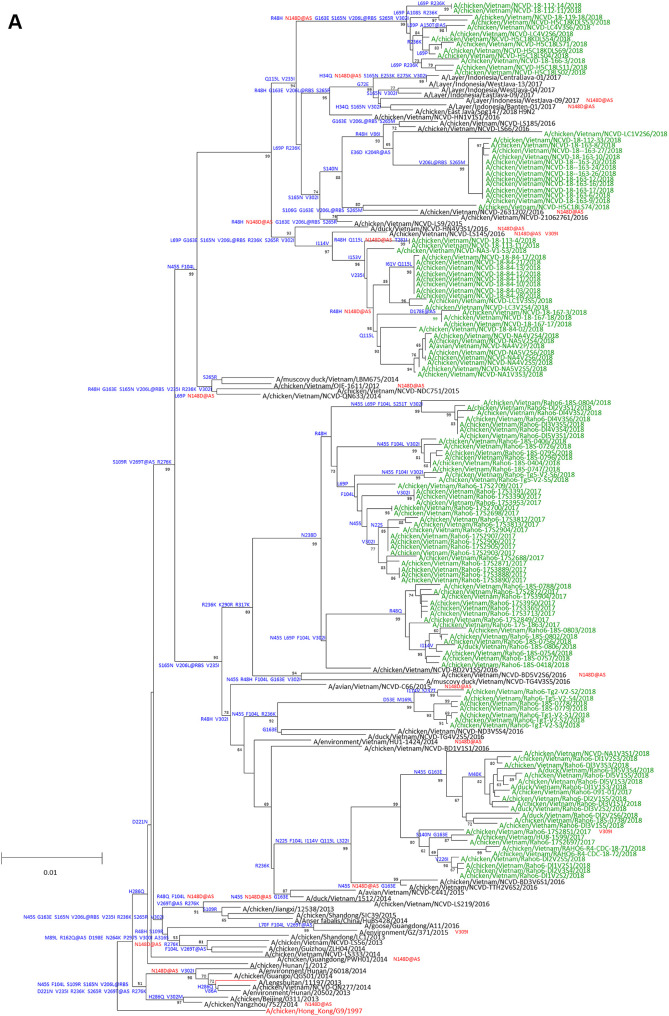
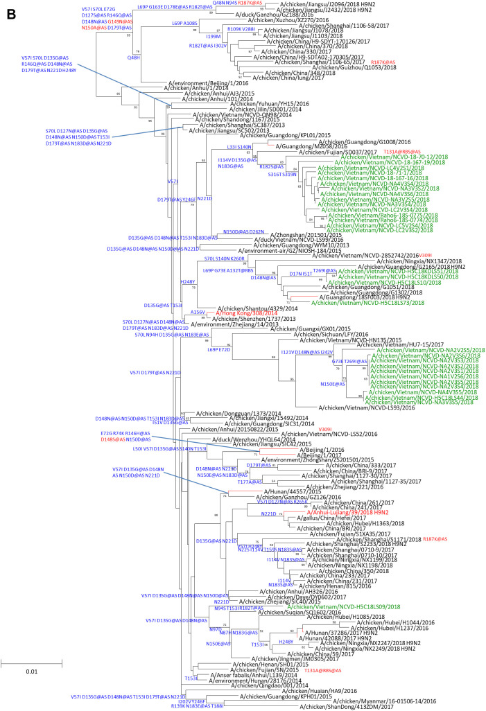
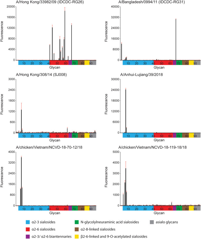
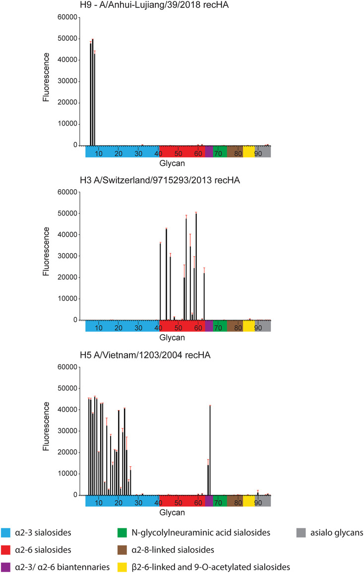
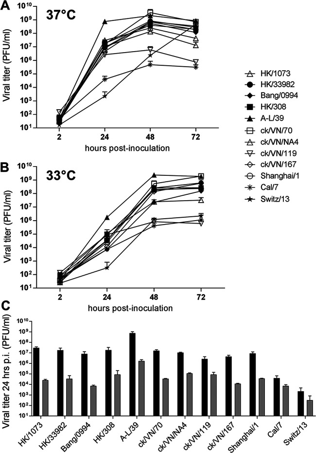
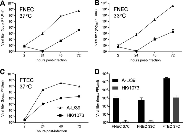
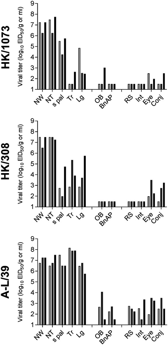
Low-pathogenicity avian influenza A(H9N2) viruses, enzootic in poultry populations in Asia, are associated with fewer confirmed human infections but higher rates of seropositivity compared to A(H5) or A(H7) subtype viruses. Cocirculation of A(H5) and A(H7) viruses leads to the generation of reassortant viruses bearing A(H9N2) internal genes with markers of mammalian adaptation, warranting continued surveillance in both avian and human populations. Here, we describe active surveillance efforts in live poultry markets in Vietnam in 2018 and compare representative viruses to G1 and Y280 lineage viruses that have infected humans. Receptor binding properties, pH thresholds for HA activation, in vitro replication in human respiratory tract cells, and in vivo mammalian pathogenicity and transmissibility were investigated. While A(H9N2) viruses from both poultry and humans exhibited features associated with mammalian adaptation, one human isolate from 2018, A/Anhui-Lujiang/39/2018, exhibited increased capacity for replication and transmission, demonstrating the pandemic potential of A(H9N2) viruses.IMPORTANCE A(H9N2) influenza viruses are widespread in poultry in many parts of the world and for over 20 years have sporadically jumped species barriers to cause human infection. As these viruses continue to diversify genetically and antigenically, it is critical to closely monitor viruses responsible for human infections, to ascertain if A(H9N2) viruses are acquiring properties that make them better suited to infect and spread among humans. In this study, we describe an active poultry surveillance system established in Vietnam to identify the scope of influenza viruses present in live bird markets and the threat they pose to human health. Assessment of a recent A(H9N2) virus isolated from an individual in China in 2018 is also reported, and it was found to exhibit properties of adaptation to humans and, importantly, it shows similarities to strains isolated from the live bird markets of Vietnam.

Keywords: H9N2; influenza; surveillance studies; transmission.


Figures

FIG 1
FIG 1
Evolution of A(H9N2) HA genes belonging to the Y280 lineages A and B. The percentages of replicate trees in which the associated taxa clustered together in the bootstrap test (1,000 replicates) are shown next to the branches. (A and B) The A Y280 (A) and B Y280 (B) viruses tested in this study are shown in green. Candidate vaccine viruses are shown in red. Amino acid differences relative to the closest candidate vaccine viruses are shown in blue on each tree branch. Mutations found in the genetic changes inventory are shown in red. Bold lettering indicates a mutation at a putative antigenic site.
FIG 1
FIG 1
Evolution of A(H9N2) HA genes belonging to the Y280 lineages A and B. The percentages of replicate trees in which the associated taxa clustered together in the bootstrap test (1,000 replicates) are shown next to the branches. (A and B) The A Y280 (A) and B Y280 (B) viruses tested in this study are shown in green. Candidate vaccine viruses are shown in red. Amino acid differences relative to the closest candidate vaccine viruses are shown in blue on each tree branch. Mutations found in the genetic changes inventory are shown in red. Bold lettering indicates a mutation at a putative antigenic site.
FIG 2
FIG 2
Glycan microarray analysis of representative A(H9N2) influenza viruses. Colored bars represent glycans that contain α-2,3 sialic acid (SA) (blue), α-2,6 SA (red), α-2,3/α-2,6 mixed SA (purple), N-glycolyl SA (green), α-2,8 SA (brown), β-2,6 and 9-O-acetyl SA (yellow), and non-SA (gray). Error bars reflect the standard error (SE) in the signal for 6 independent replicates on the array. Antisera for glycan array analysis were raised against homologous virus, with the exception of ck/VN/119 and ck/VN/70, for which antisera were raised against A-L/39 virus.
FIG 3
FIG 3
Glycan microarray analysis of recombinant HA controls. A-L/39, avian Viet04, and seasonal Switz/13 virus HAs were analyzed. Colored bars represent glycans that contain α-2,3 sialic acid (SA) (blue), α-2,6 SA (red), α-2,3/α-2,6 mixed SA (purple), N-glycolyl SA (green), α-2,8 SA (brown), β-2,6 and 9-O-acetyl SA (yellow), and non-SA (gray). Error bars reflect SE in the signal for 6 independent replicates on the array.
FIG 4
FIG 4
Replication of A(H9N2) viruses in Calu-3 cells. Human bronchial epithelial (Calu-3) cells were grown on transwell inserts and infected apically in triplicate at an MOI of 0.01 with the indicated A(H9N2), A(H7N9) (Shanghai/1), 2009 pandemic A(H1N1) (Cal/7), or seasonal A(H3N2) (Switz/13) viruses. (A and B) Cells were incubated at 37°C (A) or 33°C (B). Culture supernatants were sampled at the indicated times p.i. and titrated in MDCK cells by standard plaque assay. (C) Comparison of infectious titers collected at 24 h p.i. from 37°C (black bars) and 33°C (gray bars) cultures is shown. The limit of virus detection was 10 PFU/ml.
FIG 5
FIG 5
Replication of A(H9N2) viruses in primary ferret cell cultures. (A to C) Ferret nasal epithelial cells (FNEC) (A and B) or ferret tracheal epithelial cells (FTEC) (C) were isolated independently from three naive ferrets, grown on transwell inserts, and inoculated apically at an MOI of 0.01 with either A-L/39 or HK/1073 A(H9N2) virus. Cells were incubated at 37°C or 33°C as indicated. At each indicated time point, 200 μl of medium was added apically and collected after 10 min; all samples were titrated in MDCK cells by standard plaque assay. (D) Comparison of infectious titers collected at 24 h p.i. from all cultures is shown. The limit of virus detection was 10 PFU.
FIG 6
FIG 6
Pathogenicity of A(H9N2) viruses in ferrets. Three ferrets were inoculated intranasally with 106 EID50 of each virus indicated, and tissues and specimens were collected day 3 p.i. NW, nasal wash; NT, nasal turbinates; s pal, soft palate; Tr, trachea; Lg, lung; OB, olfactory bulb; BnAP, brain (pooled anterior and posterior); RS, rectal swab; Int, intestine (pooled duodenum, jejunoileum, and descending colon); Conj, surrounding conjunctiva. NW, NT, s pal, RS, Eye, and Conj are expressed as EID50/ml of sample or tissue homogenate, and Tr, Lg, OB, BnAP, and Int are expressed as EID50/g. The limit of virus detection was 101.5 EID50/g or ml.
FIG 7
FIG 7
Transmissibility of A(H9N2) viruses between ferrets. Six ferrets were inoculated intranasally with 106 EID50 of each virus indicated. At 24 h p.i., a naive ferret was placed in the same cage as an inoculated ferret (direct contact [DC] transmission model, left column) or in an adjacent cage with perforated side walls to an inoculated ferret (respiratory droplet [RD] transmission model, right column). Nasal washes (NW) were collected from inoculated (left sets of bars) or contact ferrets (right sets of bars) on alternate days p.i./p.c. Bars represent individual ferrets. The limit of virus detection was 101.5 EID50/ml.

References

    1. Peacock THP, James J, Sealy JE, Iqbal M. 2019. A global perspective on H9N2 avian influenza virus. Viruses 11:620. doi:10.3390/v11070620. - DOI - PMC - PubMed
    1. Li C, Wang S, Bing G, Carter RA, Wang Z, Wang J, Wang C, Wang L, Wu G, Webster RG, Wang Y, Sun H, Sun Y, Liu J, Pu J. 2017. Genetic evolution of influenza H9N2 viruses isolated from various hosts in China from 1994 to 2013. Emerg Microbes Infect 6:e106. doi:10.1038/emi.2017.94. - DOI - PMC - PubMed
    1. Pusch EA, Suarez DL. 2018. The multifaceted zoonotic risk of H9N2 avian influenza. Vet Sci 5:82. doi:10.3390/vetsci5040082. - DOI - PMC - PubMed
    1. Li X, Shi J, Guo J, Deng G, Zhang Q, Wang J, He X, Wang K, Chen J, Li Y, Fan J, Kong H, Gu C, Guan Y, Suzuki Y, Kawaoka Y, Liu L, Jiang Y, Tian G, Li Y, Bu Z, Chen H. 2014. Genetics, receptor binding property, and transmissibility in mammals of naturally isolated H9N2 avian influenza viruses. PLoS Pathog 10:e1004508. doi:10.1371/journal.ppat.1004508. - DOI - PMC - PubMed
    1. Obadan AO, Santos J, Ferreri L, Thompson AJ, Carnaccini S, Geiger G, Gonzalez Reiche AS, Rajao DS, Paulson JC, Perez DR. 2018. Flexibility in vitro of amino acid 226 in the receptor-binding site of an H9 subtype influenza A virus and its effect in vivo on virus replication, tropism, and transmission. J Virol 93:e02011-18. doi:10.1128/JVI.02011-18. - DOI - PMC - PubMed

Publication types

MeSH terms