. 2020 Jul 1;11(7):499.
doi: 10.1038/s41419-020-2704-9.

Reduced replication origin licensing selectively kills KRAS-mutant colorectal cancer cells via mitotic catastrophe

Affiliations

Reduced replication origin licensing selectively kills KRAS-mutant colorectal cancer cells via mitotic catastrophe

Bastian Gastl et al. Cell Death Dis. .

Abstract

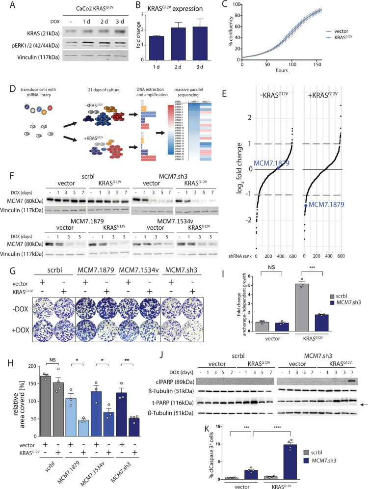
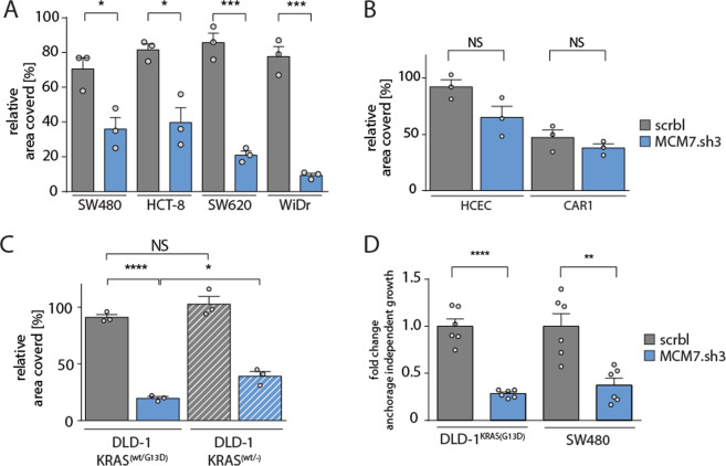
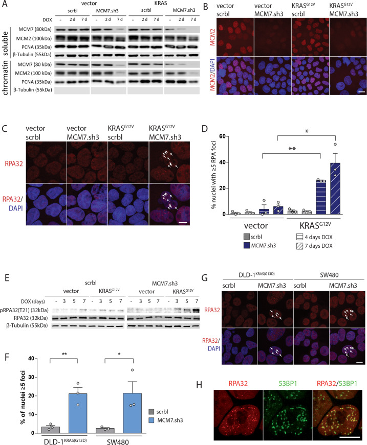
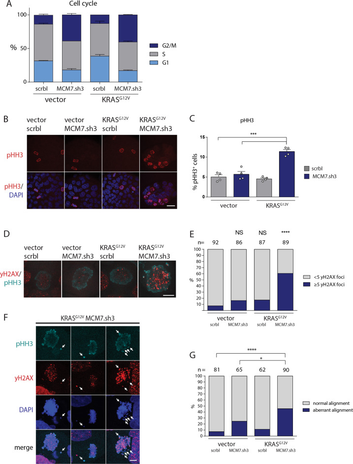
To unravel vulnerabilities of KRAS-mutant CRC cells, a shRNA-based screen specifically inhibiting MAPK pathway components and targets was performed in CaCo2 cells harboring conditional oncogenic KRASG12V. The custom-designed shRNA library comprised 121 selected genes, which were previously identified to be strongly regulated in response to MEK inhibition. The screen showed that CaCo2 cells expressing KRASG12V were sensitive to the suppression of the DNA replication licensing factor minichromosome maintenance complex component 7 (MCM7), whereas KRASwt CaCo2 cells were largely resistant to MCM7 suppression. Similar results were obtained in an isogenic DLD-1 cell culture model. Knockdown of MCM7 in a KRAS-mutant background led to replication stress as indicated by increased nuclear RPA focalization. Further investigation showed a significant increase in mitotic cells after simultaneous MCM7 knockdown and KRASG12V expression. The increased percentage of mitotic cells coincided with strongly increased DNA damage in mitosis. Taken together, the accumulation of DNA damage in mitotic cells is due to replication stress that remained unresolved, which results in mitotic catastrophe and cell death. In summary, the data show a vulnerability of KRAS-mutant cells towards suppression of MCM7 and suggest that inhibiting DNA replication licensing might be a viable strategy to target KRAS-mutant cancers.


Conflict of interest statement

The authors declare that they have no conflict of interest.

Figures

Fig. 1
Fig. 1. KRAS mutant cells are sensitive to MCM7 suppression.
a Western blot analysis shows that KRASG12V overexpression is at physiological levels and potent to induce downstream activation of ERK. Vinculin serves as a loading control. b Quantification of KRASG12V protein expression after 1, 2, and 3 days of doxycycline induction. Mean expression ± SEM is shown (n = 2–3 per group). c Representative growth curve of CaCo2 cells after KRASG12V induction shows no difference in growth with or without KRASG12V expression (n = 6 technical replicates; error bars = SEM). d Graphical outline of the RNAi screen to identify genetic interactors of mutant KRAS. e Change of shRNA abundance in the shRNA screen after 21 days of culture with and without doxycycline as determined by massive parallel sequencing. Each dot represents a shRNA. The y-axis depicts the log2 fold change of shRNA abundance. The x-axis shows each shRNA ranked by their fold change. The shRNA MCM7.1879 is specifically depleted when co-expressed with doxycycline induced KRASG12V. f Western blots showing MCM7 expression after knockdown. KRASG12V and shRNA expression were induced with doxycycline for 0, 1, 3, 5, or 7 days. MCM7 knockdown was efficient after 3 days of shRNA expression. Vinculin serves as loading control. g, h Clonogenic assays of three non-overlapping shRNAs targeting MCM7 show increased sensitivity of KRASG12V expressing CaCo2 cells to MCM7 knockdown. Mean percentage of the relative area covered by cells ± SEM is shown (n = 3–4 per group). Student’s t-test (two-sided); NS = not significant; *p < 0.05; **p < 0.01. i Soft agar assays showing anchorage-independent growth in CaCo2 cells ± induced KRASG12V and ± MCM7.sh3. KRASG12V expression increases colony formation in CaCo2 cells about five fold. MCM7 knockdown has no detectable effect on cells expressing an empty control vector, whereas cells expressing additionally KRASG12V reduce colony formation significantly. Mean fold change of colonies formed after five weeks of culture is shown ± SEM (n = 3 per group). Student’s t-test (two-sided); NS = not significant; ***p < 0.001. j Western blots show an increase in cleaved PARP (clPARP) staining after five and seven days of MCM7 knockdown specifically in CaCo2 cells expressing KRASG12V. ß-Tubulin and total PARP serve as a loading control, the cleaved PARP is indicated by an arrow. k FACS analysis of cleaved Caspase 3 (clCaspase 3) reveals a significant increase of apoptosis in cells co-expressing KRASG12V and MCM7.sh3 compared to cells expressing MCM7.sh3 alone. Mean percentage of clCaspase 3+ cells ± SEM is shown (n = 4 per group). Student’s t-test (two-sided); ***p < 0.001; ****p < 0.0001.
Fig. 2
Fig. 2. Consistent growth reduction of MCM7-suppressed colorectal cancer cell lines.
a Clonogenic assay quantification of SW480 (KRASG12V), HCT-8 (KRASG13A), HT-29 (BRAFV600E), WiDr (BRAFV600E). All cell lines showed decreased growth after doxycycline induction of MCM7.sh3 compared to the control shRNA (scrbl). Mean percentage of the relative area covered by cells ± SEM is shown (n = 3 per group). Student’s t-test (two-sided); *p < 0.05; ***p < 0.001. b Clonogenic assay quantification of DLD1KRAS(wt/−) and its parental cell line DLD1KRAS(wt/G13D). Both cell lines are sensitive to MCM7 suppression, but DLD1 cells without mutated KRAS become more resistant to low levels of MCM7. Mean percentage of the relative area covered by cells ± SEM is shown (n = 3 per group). Student’s t-test (two-sided); NS = not significant; *p < 0.05; ****p < 0.0001. c Quantification of soft agar assays confirmed decreased anchorage-independent colony formation in KRAS mutated DLD1 and SW480 cells. Mean fold change of colonies ± SEM is shown (n = 6 per group). Student’s t-test (two-sided); **p < 0.01; ****p < 0.0001.
Fig. 3
Fig. 3. Cells with MCM7 knockdown show increased replication stress after KRASG12V expression.
a Protein fractionation and subsequent western blot analysis showed a potent knockdown of MCM7 of the soluble as well as the chromatin-bound protein fraction. MCM2 levels are exclusively depleted from the chromatin-bound fraction. β-tubulin and PCNA serve as loading controls for the soluble and chromatin-bound fraction, respectively. b In situ chromatin fractionation of adherent cells followed by immunofluorescence staining shows a robust depletion of MCM2 from the chromatin after 7 days of MCM7 knockdown. Scale bar = 20 µm. c Immunofluorescence after 7 days of doxycycline induction showed RPA32 foci formation in CaCo2 cells after simultaneous KRASG12V and MCM7.sh3 expression. Arrows indicate RPA foci. Scale bar = 10 µm. d Quantification of nuclei with 5 or more RPA32 foci showed a significant increase of RPA32 focalization only when KRASG12V and MCM7.sh3 were co-expressed in CaCo2 cells. After 4 and 7 days, ∼26% and ∼40% of cells exhibited 5 or more RPA32 foci, respectively. Mean percentage of cells with 5 or more RPA32 foci ± SEM is shown (n = 3 per group). At least 50 cells per experiment were counted. Student’s t-test (two-sided); *p < 0.05, **p < 0.01. e Western blot shows specific phosphorylation of RPA32 in KRASG12V expressing CaCo2 cells after MCM7 knockdown. β-tubulin serves as a loading control. f Quantification of immunofluorescence reveals an increase of cells with RPA foci after four days of MCM7 knockdown in DLD1 and SW480 cells. Mean percentage of cells with ≥5 RPA foci ± SEM is shown (n = 3 per group). At least 50 cells per experiment were counted. Student’s t-test (two-sided); *p < 0.05; **p < 0.01. g Immunofluorescence after 4 days of doxycycline induction showed RPA32 foci formation in DLD-1 and SW480 cells after MCM7.sh3 expression. Arrows indicate RPA foci. Scale bar = 10 µm. h CaCo2 cells expressing KRASG12V and MCM7.sh3 show colocalization of RPA and 53BP1 foci after 7 days of doxycycline induction. Scale bar = 10 µm.
Fig. 4
Fig. 4. Induction of mutant KRAS expression and MCM7 suppression drives cells into mitotic catastrophe.
a Expression of KRASG12V and MCM7.sh3 in CaCo2 cells were induced with doxycycline for 7 days. BrdU was added to the cells 30 min before they were harvested and stained with propidium iodide (PI) and an anti-BrdU-AF488 antibody. FACS analysis reveals increased persistence of CaCo2 cells with low levels of MCM7 in G2/M phase independently of KRASG12V seven days after induction. (n = 2 per group). b, c Cells were stained for phospho-Histone H3 (pHH3) and counted. About 5% of all cells stained positive for pHH3 after 7 days of induction except for cells co-expressing KRASG12V and MCM7.sh3, which had an increased mitotic cell number of over 11%. Scale bar = 40 µm. Mean percentage of pHH3+ cells ± SEM is shown (n = 4 per group). At least 200 cells per experiment were counted. Student’s t-test (two-sided); ***p < 0.001. d, e Co-staining of pHH3 and γH2AX revealed that the majority of mitotic CaCo2 cells exhibit DNA damage in mitosis after co-expression of KRASG12V and MCM7.sh3, whereas KRASG12V or MCM7.sh3 expression alone, did not cause a significant increase of mitotic cells with 5 or more γH2AX foci. A total of at least 86 cells per group were counted through the course of 5 independent experiments. Fisher’s exact test; ****p < 0.0001. f, g CaCo2 cells show an increased number of cells with DNA damage and randomly distributed chromosomes throughout the cell during mitosis after 7 days of KRASG12V expression and MCM7 knockdown. Arrows indicate misaligned chromosomes. Scale bar = 10 µm. Fisher’s exact test; *p < 0.05; ****p < 0.0001.

Similar articles

Cited by

References

    1. Cox AD, Fesik SW, Kimmelman AC, Luo J, Der CJ. Drugging the undruggable RAS: mission possible? Nat. Rev. Drug Disco. 2014;13:828–851. - PMC - PubMed
    1. Papke B, Der CJ. Drugging RAS: know the enemy. Science. 2017;355:1158–1163. - PubMed
    1. Ostrem JM, Peters U, Sos ML, Wells JA, Shokat KM. K-Ras(G12C) inhibitors allosterically control GTP affinity and effector interactions. Nature. 2013;503:548–551. - PMC - PubMed
    1. Janes MR, et al. Targeting KRAS mutant cancers with a covalent G12C-specific inhibitor. Cell. 2018;172:578–589.e17. - PubMed
    1. Pylayeva-Gupta. Y, Grabocka E, Bar-Sagi D. RAS oncogenes: weaving a tumorigenic web. Nat. Rev. Cancer. 2011;11:761–774. - PMC - PubMed

Publication types

Substances