. 2020 Jul 1;10(1):10729.
doi: 10.1038/s41598-020-67723-x.

Injury intensifies T cell mediated graft-versus-host disease in a humanized model of traumatic brain injury

Affiliations

Injury intensifies T cell mediated graft-versus-host disease in a humanized model of traumatic brain injury

Miguel F Diaz et al. Sci Rep. .

Abstract

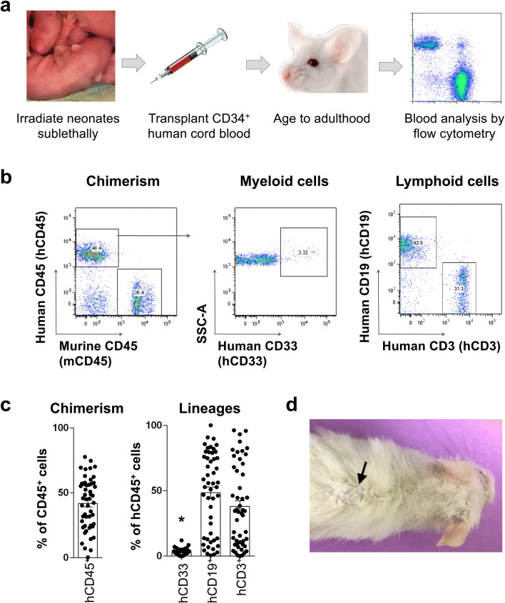
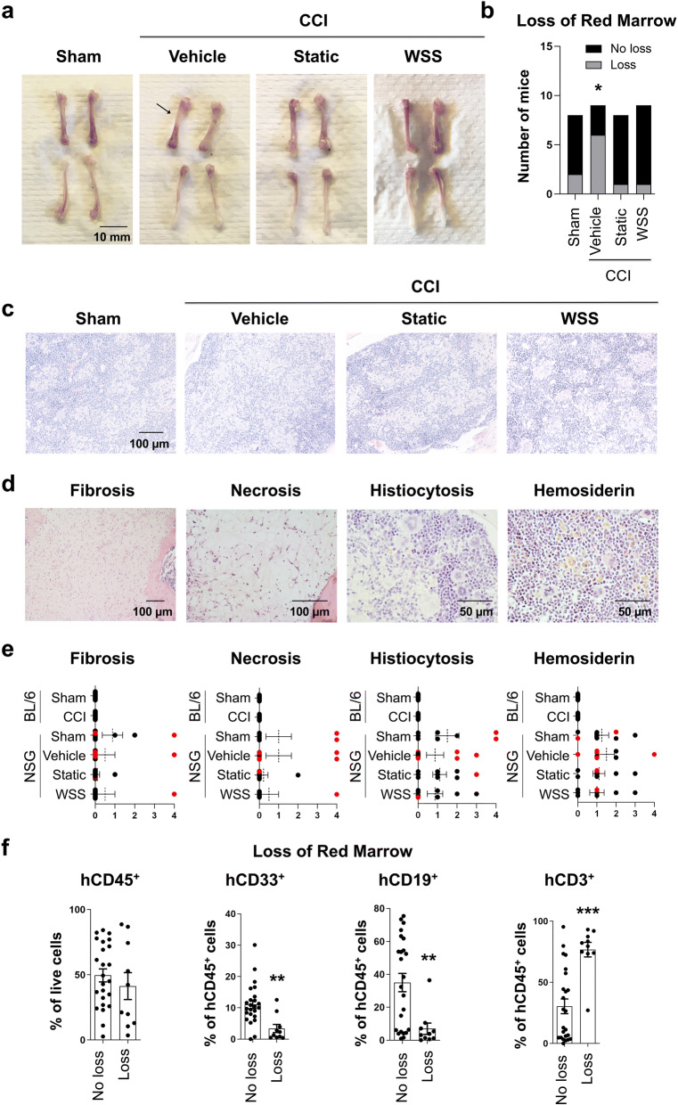
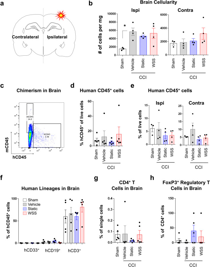
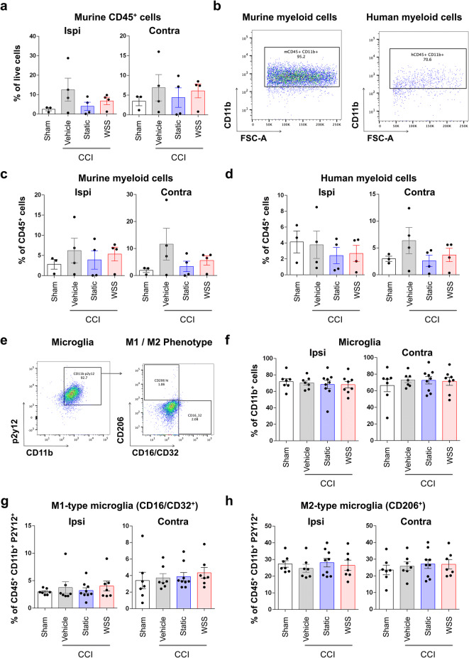
The immune system plays critical roles in promoting tissue repair during recovery from neurotrauma but is also responsible for unchecked inflammation that causes neuronal cell death, systemic stress, and lethal immunodepression. Understanding the immune response to neurotrauma is an urgent priority, yet current models of traumatic brain injury (TBI) inadequately recapitulate the human immune response. Here, we report the first description of a humanized model of TBI and show that TBI places significant stress on the bone marrow. Hematopoietic cells of the marrow are regionally decimated, with evidence pointing to exacerbation of underlying graft-versus-host disease (GVHD) linked to presence of human T cells in the marrow. Despite complexities of the humanized mouse, marrow aplasia caused by TBI could be alleviated by cell therapy with human bone marrow mesenchymal stromal cells (MSCs). We conclude that MSCs could be used to ameliorate syndromes triggered by hypercytokinemia in settings of secondary inflammatory stimulus that upset marrow homeostasis such as TBI. More broadly, this study highlights the importance of understanding how underlying immune disorders including immunodepression, autoimmunity, and GVHD might be intensified by injury.


Conflict of interest statement

C.S.C. serves on the Scientific Advisory Board for CBR, Inc. and conducts sponsored research with Athersys, Inc., Celgene Therapeutics, Inc., and CBR. K.R.A, B.S.G., C.S.C., and P.L.W. are inventors on a patent for conditioning of stem and progenitor cells for cellular therapy (U.S. Patent US20180187141A1). All other authors declare no conflict of interest.

Figures

Figure 1
Figure 1
Human T and B cells predominate in grafts from human cord blood. (a) Immunocompromised NSG mice were transplanted with CD34+-enriched human cord blood as neonates via facial vein and monitored for human chimerism in the peripheral blood. (b, c) Human and mouse leukocytes were distinguished in the peripheral blood by surface expression of human CD45 and murine CD45. Blood lineages were detected with human-specific antibodies to CD33 (myeloid), CD19 (B cells), and CD3 (T cells). Analysis revealed robust contribution of CD34+ cells to T and B cells but poor reconstitution of myeloid cells in the peripheral blood (Kruskal–Wallis one-way ANOVA with Tukey test, *p < 0.05). Data points represent 50 individual mice at 2–5 months of age, and error bars indicate standard error of the mean (SEM). (d) Photograph of a humanized mouse reveals large patches of white, flaking skin apparent on the back (arrow) and head.
Figure 2
Figure 2
Human chimerism is reduced after CCI. (a) Traumatic brain injury (TBI) was administered by CCI to the right parietal association cortex between bregma and lambda. Mice received intravenous vehicle or MSCs cultured under static conditions or preconditioned transiently with 3 h exposure to WSS. Tissues were collected 7 days after injury. (b, c) Peripheral blood was analyzed for surface expression of mCD45 and hCD45 7 days after injury, along with human lineage markers. (d) Frequency of hCD45+ cells is decreased in the periphery after injury (One-way ANOVA with Holm-Sidak test, *p = 0.02). Pre-injury chimerism from 1–3 months before injury was used to calculate percent change. Lymphocyte production shifted with age from hCD19+ B cells to greater numbers of CD3+ T cells in all treatment groups during the course of 1–3 months. Data from 8–9 individual mice per group are shown, and SEM is indicated by error bars.
Figure 3
Figure 3
Spleen size correlates with human T cell chimerism. (a) Gross examination of spleens was conducted 7 days after surgery. (b) Measurement of spleen length of 7–8 mice per group reveals no significant reduction in size after injury (30 mice total). (c) Instead, spleen length was associated with frequency of hCD45+ and hCD3+ cells in the spleen and peripheral blood (Regression analysis, p < 0.05 for all comparisons). (d) No relationship exists between treatment group and human chimerism in the spleen or inguinal lymph nodes (8–9 mice per group for analysis of spleen and 4–5 mice per group for lymph nodes).
Figure 4
Figure 4
Brain injury is associated with bone marrow aplasia. (a) Evidence for loss of red marrow was apparent in several femurs collected from humanized mice 7 days after injury. Arrow indicates clearance of marrow in femur from vehicle control. (b) Graph represents quantification of mice with signs of bone marrow clearance from 8–9 mice per group (Chi-square test, *p = 0.03). (c) Histopathological examination revealed diffuse aggregates of histiocytes within the marrow of several animals, visible as eosinophilic areas among densely packed nucleated cells. Representative photomicrographs of femurs from each treatment group are shown. (d) Examples of necrosis, histiocytosis, and hemosiderin (brown iron deposits) are displayed. (e) Histopathological scores were based upon an ordinal numeric scoring system wherein 0 represented no abnormality and 4 displayed severe signs of pathology. NSG groups included 8–9 mice per treatment, and C57BL/6 mice included 9 mice per group. Red points represent mice scored as having loss of red marrow by gross observation. (f) Frequency of hCD3+ T cells in the bone marrow correlated with red marrow destruction (Mann–Whitney rank sum test, ***p < 0.001). Frequencies of hCD33+ myeloid or hCD19+ B cells were low in the bone marrow of mice with red marrow loss in one or both femurs as determined by gross observation (Mann–Whitney rank sum test, **p < 0.003). A total of 34 mice were included in these analyses and are plotted as individual points, along with mean and SEM.
Figure 5
Figure 5
Cell therapy with MSCs elevates regulatory T cell frequencies in the bone marrow and lymph nodes. (a) Human CD4+ and regulatory T cells were measured before and 7 days after injury. Regulatory T cells were identified by CD4, CD25, and intracellular expression of FOXP3. (b) Frequency of hCD4+ T cells and Treg were quantified in the peripheral blood of 16 mice before assignment to a treatment group. (c) After injury, hCD4+ T cell frequency was generally lower but increased in the lymph nodes by therapy with MSCs preconditioned by WSS. 8–9 mice were analyzed per group. (d) Tregs were reduced in multiple hematopoietic organs after neurotrauma but increased after therapy with MSCs (n = 8–9 mice per group).
Figure 6
Figure 6
Human CD3+ T cells predominate in the brain. (a) Ipsilateral (injured) and contralateral (uninjured) hemispheres of the brain were processed for separate analysis. (b) Total cellularity of the ipsilateral hemisphere was increased by injury (One-way ANOVA with Holm-Sidak test, *p = 0.04). MSC therapy did not suppress expansion in cell numbers. Analysis of separate hemispheres included 3–4 mice in each group. (c) Human chimerism of dissociated brain tissue was assessed by flow cytometry. (d, e) Frequency of human CD45+ cells in total brain tissue (both hemispheres, n = 5 mice per group) and in separately processed hemispheres (n = 3–4 mice per group) is unaltered by injury. (f) Across all treatment groups, a small fraction of human myeloid or B cells were detected in the brain; whereas, T cells accounted for up to 100% of hCD45+ cells in several mice. (g) Injury appears to reduce CD4+ T cell frequency in the brain, though high variability exists between mice. (h) The fraction of human CD4+ T cells identifiable as FoxP3+ regulatory T cells is variable across treatment groups.
Figure 7
Figure 7
M1–M2-type microglial frequencies are not detectably altered 7 days after injury. (a) No significant change in murine CD45+ cells was observed in the brain. Hemispheres were evaluated separately in 3–4 mice per group. (b) Myeloid cells were detected by human and mouse reactive CD11b antibody. (c, d) Frequencies of mCD45+ CD11b+ and hCD45+ CD11b+ cells in the brain were not significantly different between treatment groups. (e) CD45+ CD11b+ P2Y12+ microglia were characterized as alternatively activated CD206+ M2-type microglia or inflammatory CD16/CD32+ M1-type microglia. (f) Frequencies of microglia in the ipsilateral and contralateral hemispheres appear unaffected after injury. Microglia were evaluated in 7–9 mice per group. (g, h) M1-type and M2-type microglia labeled by surface detection of CD16/CD32 and CD206, respectively, are not noticeably altered after injury.

References

    1. McDonald SJ, Sun M, Agoston DV, Shultz SR. The effect of concomitant peripheral injury on traumatic brain injury pathobiology and outcome. J. Neuroinflamm. 2016;13:90. - PMC - PubMed
    1. Xiong Y, Mahmood A, Chopp M. Animal models of traumatic brain injury. Nat. Rev. Neurosci. 2013;14:128–142. - PMC - PubMed
    1. Brooks GA, Martin NA. Cerebral metabolism following traumatic brain injury: New discoveries with implications for treatment. Front. Neurosci. 2014;8:408. - PMC - PubMed
    1. Fuchs A, et al. Trauma induces emergency hematopoiesis through IL-1/MyD88-dependent production of G-CSF. J. Immunol. 2019;202:3020. - PMC - PubMed
    1. Livingston DH, et al. Bone marrow failure following severe injury in humans. Ann. Surg. 2003;238:748–753. - PMC - PubMed

Publication types