Review
. 2020 Jun 16:14:569.
doi: 10.3389/fnins.2020.00569. eCollection 2020.

One Is Not Enough: Understanding and Modeling Polysubstance Use

Affiliations
Review

One Is Not Enough: Understanding and Modeling Polysubstance Use

Elizabeth A Crummy et al. Front Neurosci. .

Abstract

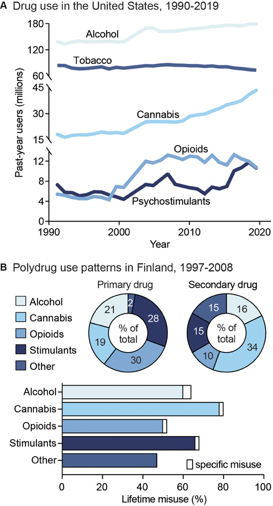
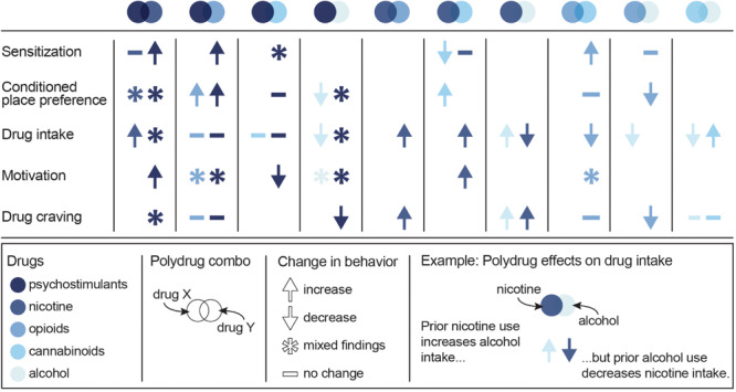
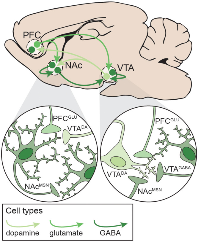
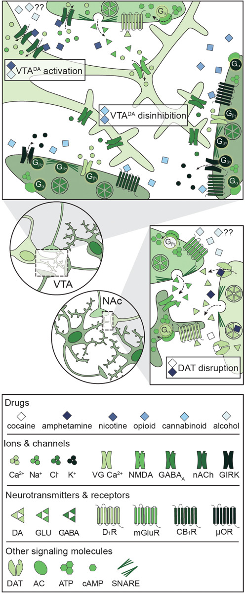
Substance use disorder (SUD) is a chronic, relapsing disease with a highly multifaceted pathology that includes (but is not limited to) sensitivity to drug-associated cues, negative affect, and motivation to maintain drug consumption. SUDs are highly prevalent, with 35 million people meeting criteria for SUD. While drug use and addiction are highly studied, most investigations of SUDs examine drug use in isolation, rather than in the more prevalent context of comorbid substance histories. Indeed, 11.3% of individuals diagnosed with a SUD have concurrent alcohol and illicit drug use disorders. Furthermore, having a SUD with one substance increases susceptibility to developing dependence on additional substances. For example, the increased risk of developing heroin dependence is twofold for alcohol misusers, threefold for cannabis users, 15-fold for cocaine users, and 40-fold for prescription misusers. Given the prevalence and risk associated with polysubstance use and current public health crises, examining these disorders through the lens of co-use is essential for translatability and improved treatment efficacy. The escalating economic and social costs and continued rise in drug use has spurred interest in developing preclinical models that effectively model this phenomenon. Here, we review the current state of the field in understanding the behavioral and neural circuitry in the context of co-use with common pairings of alcohol, nicotine, cannabis, and other addictive substances. Moreover, we outline key considerations when developing polysubstance models, including challenges to developing preclinical models to provide insights and improve treatment outcomes.

Keywords: addiction; neurobiology of addiction; neuronal signaling and behavior; polydrug; preclinical models; review; reward circuitry; substance use.


Figures

FIGURE 1
FIGURE 1
Public health trends in drug use. (A) Drug use in the United States from 1990 to 2019. Data from the National Household Survey on Drug Abuse (Substance Abuse and Mental Health Services Administration, 1993; Substance Abuse and Mental Health Services Administration, 1995; Substance Abuse and Mental Health Services Administration, 1997; Substance Abuse and Mental Health Services Administration, 2003) and the National Survey on Drug Use and Health (Substance Abuse and Mental Health Services Administration, 2005; Substance Abuse and Mental Health Services Administration, 2007; Substance Abuse and Mental Health Services Administration, 2009; Substance Abuse and Mental Health Services Administration, 2011; Substance Abuse and Mental Health Services Administration, 2013; Substance Abuse and Mental Health Services Administration, 2015; Substance Abuse and Mental Health Services Administration, 2017; Substance Abuse and Mental Health Services Administration, 2019). (B) Unspecified polysubstance use in treatment-seeking drug users in Finland from 1997 to 2008. Top: Primary (left) and secondary (right) drugs used by treatment-seeking drug users, shown as percent of total users. Bottom: Percent of users reporting exclusive misuse of one drug (white bars) or misuse of a given drug along with polysubstance use of another (colored bars). Data from Onyeka et al. (2012).
FIGURE 2
FIGURE 2
Summary of the effects of specific polydrug combinations on assays of addiction-like behaviors. Studies are organized into X/Y polydrug combos (columns) and behavioral assays (rows), with subcolumns for the effect of drug Y on drug X (left subcolumn) and the effect of drug X on drug Y (right subcolumn). Symbols represent the net effect of the X/Y polydrug combo on a given behavior, with color depicting the specific drugs tested. Sensitization: locomotor sensitization; Conditioned place preference: acquisition, expression; Drug intake: self-administration; Motivation: progressive ratio, behavioral economics; Drug craving: reinstatement, cue reactivity. Data from Mello and Mendelson (1978), Mello et al. (1980), Mello et al. (2014), Huston-Lyons et al. (1993), Foltin et al. (1993), Aspen and Winger (1997), Ranaldi and Wise (2000), Valverde et al. (2001); De Vries et al. (2001), Parker et al. (2004); Solinas et al. (2005), Liang et al. (2006); Biala and Budzynska (2006), Ward et al. (2006); Panlilio et al. (2007), Panlilio et al. (2013), Winger et al. (2007), Lê et al. (2010), Lê et al. (2014), Levine et al. (2011), Cortright et al. (2011), Pomfrey et al. (2015), Maguire and France (2016); Mahmud et al. (2017), Fredriksson et al. (2017); Giasson-Gariépy et al. (2017), Griffin et al. (2017); Schwartz et al. (2018), Winkler et al. (2018); Manwell et al. (2019), Ponzoni et al. (2019), Crummy et al. (2020), and Stennett et al. (2020).
FIGURE 3
FIGURE 3
Neural circuitry targeted by potentially addictive drugs. Simplified schematic emphasizing local and distal connections between the PFC, NAc, and VTA that are targeted by potentially addictive drugs. Left: NAcMSNs receive excitatory glutamatergic inputs from PFCGLU neurons, dopaminergic inputs from VTADA neurons, and inhibitory GABAergic inputs from other NAcMSNs. Right: VTADA neurons are maintained under tonic inhibition by local VTAGABA interneurons and NAcMSNs and receive excitatory inputs from PFCGLU neurons. DA: dopamine; GLU: glutamate; MSN: medium spiny neuron; NAc: nucleus accumbens; PFC: prefrontal cortex; VTA: ventral tegmental area.
FIGURE 4
FIGURE 4
Primary mechanisms of action of potentially addictive drugs. Potentially addictive drugs increase DA release into the NAc, but different drugs act via distinct mechanisms. Top: Opioids and cannabinoids disinhibit VTADA neurons via presynaptic inhibition of VTAGABA and NAcMSN inputs through four notable mechanisms: Inhibition of VG Ca2+ channels, activation of GIRKs, inactivation of AC, and inhibition of GABA release. Nicotine activates VTADA neurons via direct activation of somatodendritic nAChRs and activation of presynaptic PFCGLU inputs. Alcohol directly activates VTADA cell bodies, but the mechanism is not understood. Bottom: Psychostimulants impair DA reuptake by blocking DAT (cocaine) or reversing the activity of DAT and facilitating DA release (amphetamine), leading to increased dopaminergic tone in the NAc. Alcohol also targets PFCGLU inputs to the NAc, but the net effect on PFCGLU activity is unknown. AC: adenylyl cyclase; ATP: adenosine triphosphate; cAMP: cyclic adenosine monophosphate; CB1R: cannabinoid 1 receptor; DA: dopamine; DAT: dopamine transporter; D1R: dopamine D1 receptor; GABA: gamma- aminobutyric acid; GIRK: G protein-coupled inwardly rectifying K+ channel; GLU: glutamate; mGluR: metabotropic glutamate receptor; μOR: mu opioid receptor; NAc: nucleus accumbens; nACh: nicotinic acetylcholine receptor; NMDA: N-methyl-D-aspartate receptor; PFC: prefrontal cortex; SNARE: soluble N-ethylmaleimide-sensitive factor attachment protein receptor; VG Ca2+: voltage-gated Ca2+ channel; VTA: ventral tegmental area.
FIGURE 5
FIGURE 5
Persistent disruptions in synaptic and structural plasticity caused by long-term use of potentially addictive drugs. Studies are organized by cell type (columns) and type of disruption (rows), with symbols depicting the net change in plasticity and color depicting which drug was tested. Data from Bonci and Williams (1996), Kang et al. (1996, 1998), Robinson and Kolb (1997), Robinson and Kolb (2004), Badiani et al. (1999), Mansvelder and McGehee (2000), Dahchour and De Witte (2000), Uslaner et al. (2001), Brown and Kolb (2001), Robinson et al. (2002), Saal et al. (2003), Amantea et al. (2004), Hamilton and Kolb (2005), Nasif et al. (2005), Kolb et al. (2006), Kolb et al. (2018), Huang et al. (2007), Zhou et al. (2007), Van Den Oever et al. (2008), Kalivas et al. (2009), Niehaus et al. (2010), Russo et al. (2010), Bowers et al. (2010), Spiga et al. (2010), Levine et al. (2011), Dacher and Nugent (2011), Kroener et al. (2012), Lobo et al. (2013), Trifilieff and Martinez (2013), Mello et al. (2014), Peterson et al. (2015), Bloomfield et al. (2016), Creed et al. (2016), Ehlinger et al. (2016), Hearing et al. (2016), Morud et al. (2016), Friend L. et al., 2017), Langlois and Nugent (2017), Edwards et al. (2017), Spencer et al. (2018), You et al. (2018), Hwang and Lupica (2019), Kruse et al. (2019), McDevitt et al. (2019), Neuhofer et al. (2019), Pickel et al. (2019), and Ponzoni et al. (2019).

References

    1. Aguayo L. G. (1990). Ethanol potentiates the GABAA-activated Cl- current in mouse hippocampal and cortical neurons. Eur. J. Pharmacol. 187 127–130. 10.1016/0014-2999(90)90349-B - DOI - PubMed
    1. Aharonovich E., Liu X., Samet S., Nunes E., Waxman R., Hasin D. (2005). Postdischarge cannabis use and its relationship to cocaine, alcohol, and heroin use: a prospective study. Am. J. Psychiatry 162 1507–1514. 10.1176/appi.ajp.162.8.1507 - DOI - PubMed
    1. Al-Hasani R., Bruchas M. R. (2011). Molecular mechanisms of opioid receptor-dependent signaling and behavior. Anesthesiology 115 1363–1381. 10.1097/ALN.0b013e318238bba6 - DOI - PMC - PubMed
    1. Amantea D., Tessari M., Bowery N. G. (2004). Reduced G-protein coupling to the GABAB receptor in the nucleus accumbens and the medial prefrontal cortex of the rat after chronic treatment with nicotine. Neurosci. Lett. 355 161–164. 10.1016/j.neulet.2003.10.060 - DOI - PubMed
    1. Arvidsson U., Riedl M., Chakrabarti S., Lee J. H., Nakano A. H., Dado R. J., et al. (1995). Distribution and targeting of a μ-opioid receptor (MOR1) in brain and spinal cord. J. Neurosci. 15 3328–3341. 10.1523/jneurosci.15-05-03328.1995 - DOI - PMC - PubMed