. 2020 Oct;36(10):2409-2425.
doi: 10.1007/s00381-020-04775-4. Epub 2020 Jul 1.

Management of spinal deformities and tibial pseudarthrosis in children with neurofibromatosis type 1 (NF-1)

Affiliations

Management of spinal deformities and tibial pseudarthrosis in children with neurofibromatosis type 1 (NF-1)

Kiril V Mladenov et al. Childs Nerv Syst. 2020 Oct.

Erratum in

Abstract

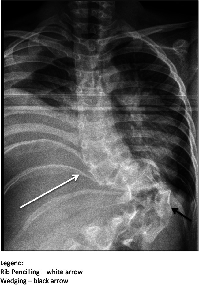
The skeletal system is affected in up to 60% of patients with neurofibromatosis type 1. The most commonly observed entities are spinal deformities and tibial dysplasia. Early recognition of radiologic osseous dystrophy signs is of utmost importance because worsening of the deformities without treatment is commonly observed and surgical intervention is often necessary. Due to the relative rarity and the heterogenic presentation of the disease, evidence regarding the best surgical strategy is still lacking.

Purpose: To report our experience with the treatment of skeletal manifestations in pediatric patients with (neurofibromatosis type 1) NF-1 and to present the results with our treatment protocols.

Materials and methods: This is a retrospective, single expert center study on children with spinal deformities and tibial dysplasia associated with NF-1 treated between 2006 and 2020 in a tertiary referral institution.

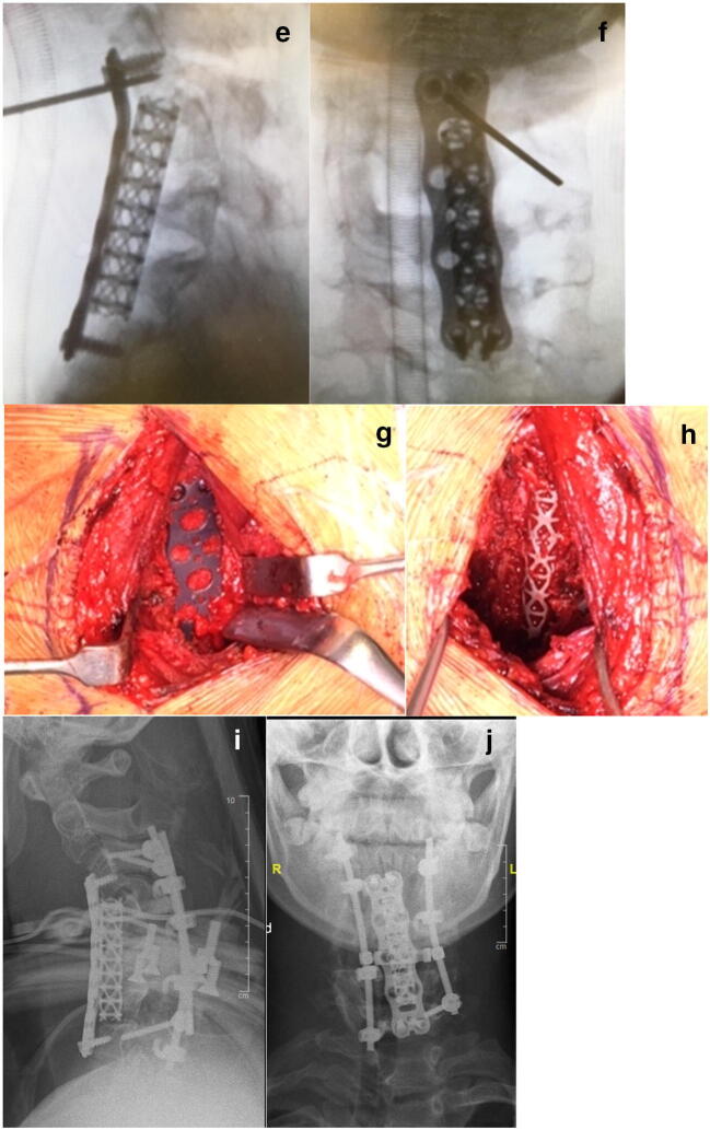
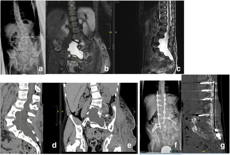
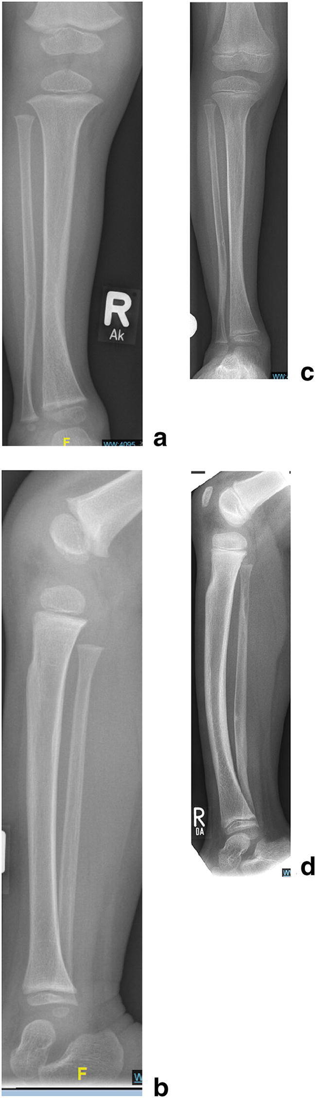
Results: Spinal deformity: Thirty-three patients (n = 33) were included. Mean age at index surgery was 9.8 years. In 30 patients (91%), the deformity was localized in the thoracic and/or lumbar spine, and in 3 patients (9%), there was isolated involvement of the cervical spine. Eleven patients (33%) received definitive spinal fusion as an index procedure and 22 (67%) were treated by means of "growth-preserving" spinal surgery. Halo-gravity traction before index surgery was applied in 11 patients (33%). Progression of deformity was stopped in all patients and a mean curve correction of 60% (range 23-98%) was achieved. Mechanical problems with instrumentation requiring revision surgery were observed in 55% of the patients treated by growth-preserving techniques and in none of the patients treated by definitive fusion. One patient (3%) developed a late incomplete paraplegia due to a progressive kyphotic deformity. Tibial dysplasia: The study group comprised of 14 patients. In 5 of them (36%) pathological fractures were present on initial presentation. In the remaining 9 patients (64%), anterior tibial bowing without fracture was observed initially. Four of them (n = 4, 28%) subsequently developed a pathologic fracture despite brace treatment. Surgical treatment was indicated in 89% of the children with pathological fractures. This involved resection of the pseudarthrosis, autologous bone grafting, and intramedullary nailing combined with external fixation in some of the cases. In 50% of the patients, bone morphogenic protein was used "off-label" in order to promote union. Healing of the pseudarthrosis was achieved in all of the cases and occurred between 5 to 13 months after the index surgical intervention. Four of the patients treated surgically needed more than one surgical intervention in order to achieve union; one patient had a re-fracture. All patients had a good functional result at last follow-up.

Conclusion: Early surgical intervention is recommended for the treatment dystrophic spinal deformity in children with NF-1. Good and sustainable curve correction without relevant thoracic growth inhibition can be achieved with growth-preserving techniques alone or in combination with short spinal fusion at the apex of the curve. Preoperative halo-gravity traction is a safe and very effective tool for the correction of severe and rigid deformity in order to avoid neurologic injury. Fracture union in tibial dysplasia with satisfactory functional results can be obtained in over 80% of the children by means of surgical resection of the pseudarthrosis, intramedullary nailing, and bone grafting. Wearing a brace until skeletal maturity is achieved is mandatory in order to minimize the risk of re-fracture.

Keywords: Congenital pseudarthrosis of the tibia; Dystrophic scoliosis; Neurofibromatosis type 1.


Conflict of interest statement

On behalf of all authors, the corresponding author states that there is no conflict of interest.

Figures

Fig. 1
Fig. 1
(Case 1) Lateral MRI (a) and plain X-ray films (b) of a 15-year-old patient with NF-1 showing severe kyphosis of the C-spine. Clinical image in preoperative halo-gravity traction (c). X-rays 2 weeks and 4 weeks (d) after the beginning of the HGT. Intraoperative image intensifier images after anterior cage and plate insertion (e, f) and clinical photos of the surgical site (g, h). Postoperative X-rays in a halo jacket (i, j)
Fig. 1
Fig. 1
(Case 1) Lateral MRI (a) and plain X-ray films (b) of a 15-year-old patient with NF-1 showing severe kyphosis of the C-spine. Clinical image in preoperative halo-gravity traction (c). X-rays 2 weeks and 4 weeks (d) after the beginning of the HGT. Intraoperative image intensifier images after anterior cage and plate insertion (e, f) and clinical photos of the surgical site (g, h). Postoperative X-rays in a halo jacket (i, j)
Fig. 2
Fig. 2
(Case 2) Eight-year-old patient with an “idiopathic-like” early-onset scoliosis (ad). The curve showed mild progression to 27° (a). Brace treatment was initiated and good curve correction could be achieved (b). Full-time brace wear was recommended and further worsening of the scoliosis was prevented until skeletal maturity was reached. The brace treatment was discontinued at age 15 (c). One year after brace discontinuation: the curve remained unchanged (d)
Fig. 3
Fig. 3
(Case 3) A nine-year-old NF-1 patient with progressive dystrophic scoliosis (a, b). A short segment anterior apical fusion (T8–11) was performed to correct apical deformity and achieve stable union (a). In a second surgery, growth-preserving, vertebra-based MCGR instrumentation (T3–L3) was used in order to correct residual long segment scoliosis and global kyphosis (b)
Fig. 4
Fig. 4
(Case 4) Dystrophic scoliosis of 71° in a 15-year-old patient with NF-1 (a, b). The curve was flexible on side bending. The patient was treated by posterior spinal fusion T2–L2 with good results on latest follow-up 3 years after initial surgery (c, d).
Fig. 5
Fig. 5
(Case 5) Rib head protrusion into the spinal canal (arrow)
Fig. 6
Fig. 6
Dystrophic lumbar scoliosis (ag) in a 13-year-old NF-1 patient (a). Notice the severe dural ectasia visible on MRI (b, c) as well as the severe bone erosion resulting in a poor bone stock visible on the preoperative CT scan (d, e).Plain X-ray and noncontrast CT scan after combined anterior and posterior spinal fusion with anterior mesh cages and “off-label” rhBMP-2 showing good correction and stable fusion on latest follow-up (f, g)
Fig. 7
Fig. 7
(Case 7) Eleven-year-old NF-1 patient with moderate dystrophic scoliosis (al) . The index procedure comprised a growth-preserving technique by means of vertical expandable titanium ribs (VEPTR). The curve was well-controlled and worsening was prevented for 4 years after initial surgery (a, b). During the pubertal growth spurt, significant curve progression occurred. An “in situ” posterior fusion combined with concave instrumentation was performed at age 13 (c, d). On further follow-up, the curve increased significantly due to the “modulation” process of dystrophic alterations (e, f). Note the severe dystrophic alterations and the complexity of the deformity seen on reconstruction CT scans (g, h). The patient developed an incomplete flaccid paraplegia of the lower extremities due to an increase of thoracic kyphosis resulting in anterior compression of the spinal cord. The treatment approach comprised of removal of instrumentation and halo-gravity traction for 4 weeks (h1) followed by posterior and anterior spinal fusion. A photo of the anterior procedure showing the intervertebral cages and the strut rib graft in place (j). Postoperative X-rays showing good deformity correction (k, l)
Fig. 8
Fig. 8
(Case 8) Tibial dysplasia (ad). Tibial bowing in a 2-year-old child with tibial dysplasia (a, b). Brace management was prescribed. Follow-up after 3 years. The lateral view shows minor progression of anterior bowing but no significant worsening of the dysplastic changes with good intramedullary-cortical differentiation (c, d). Further brace wear was recommended
Fig. 9
Fig. 9
(Case 9) Antero-lateral tibial bowing (ao) in a 3-year-old child (a, b). Pathological fracture at age 4.5 years despite bracing (c, d). The extent of dystrophic changes is seen on MRI (e, f). Resection of the pseudarthrosis, retrograde intramedullary nailing and application of a ring fixator for segment bone transport in order to fill the defect (g, h). Bone segment transport with concurrent distraction osteogenesis(i, j). Six weeks after the “Docking procedure” after completion of the bone segment transport with telescopic intramedullary nail osteosynthesis and rhBMP-2 application. The external ring fixator is already removed (k, l). Final result with solid union a.p. (m), lateral (n), CT scan (o). The intramedullary telescopic nail is retained, the child should wear a protective “double shell” brace until maturity.
Fig. 10
Fig. 10
Typical appearance of a short-segment dystrophic scoliosis in a patient with NF-1
Fig. 11
Fig. 11
Typical appearance of posterior vertebral scalopping

References

    1. Akbarnia BA, Gabriel KR, Beckman E, Chalk D. Prevalence of scoliosis in neurofibromatosis. Spine. 1992;17:S244–S248. doi: 10.1097/00007632-199208001-00005. - DOI - PubMed
    1. Funasaki H, Winter RB, Lonstein JB. Denis F pathophysiology of spinal deformities in neurofibromatosis. J Bone Joint Surg Am. 1994;76A:692–700. doi: 10.2106/00004623-199405000-00010. - DOI - PubMed
    1. Lykissas MG, Schorry EK, Crawford AH, Gaines S, Rieley M, Jain VV. Does the presence of dystrophic features in patients with type 1 neurofibromatosis and spinal deformities increase the risk of surgery? Spine (Phila Pa 1976) 2013;38(18):1595–1601. doi: 10.1097/BRS.0b013e31829a7779. - DOI - PubMed
    1. Young-Hing K, Kalamchi A, MacEwen GD. Cervical spine abnormalities in neurofibromatosis. J Bone Joint Surg Am. 1979;61:659–659. - PubMed
    1. Yifei G, Xiaolong S, Yang L, Peng C, Wen Y. Clinical outcomes of anterior correction and reconstruction for neurofibromatosis-associated severe cervical kyphotic deformity. Int Orthop. 2019;43(3):639–646. doi: 10.1007/s00264-018-4050-4. - DOI - PubMed