. 2020 Aug;34(8):10731-10750.
doi: 10.1096/fj.202000671R. Epub 2020 Jul 2.

Trafficking of cholesterol from lipid droplets to mitochondria in bovine luteal cells: Acute control of progesterone synthesis

Affiliations

Trafficking of cholesterol from lipid droplets to mitochondria in bovine luteal cells: Acute control of progesterone synthesis

Michele R Plewes et al. FASEB J. 2020 Aug.

Abstract

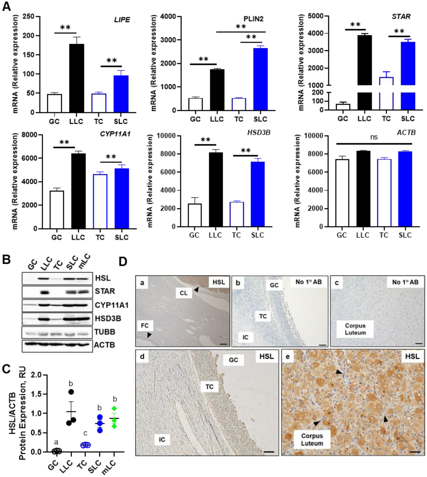
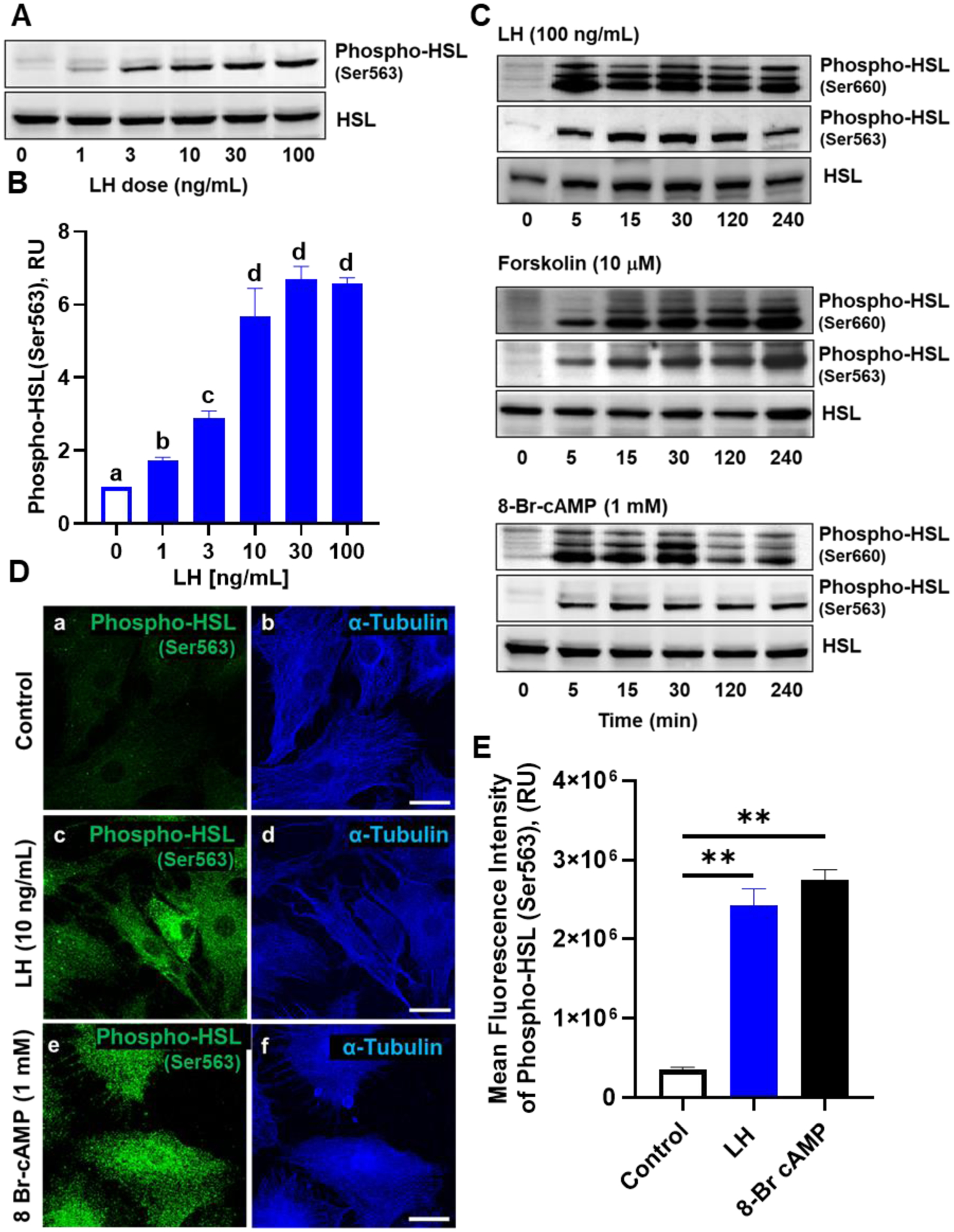
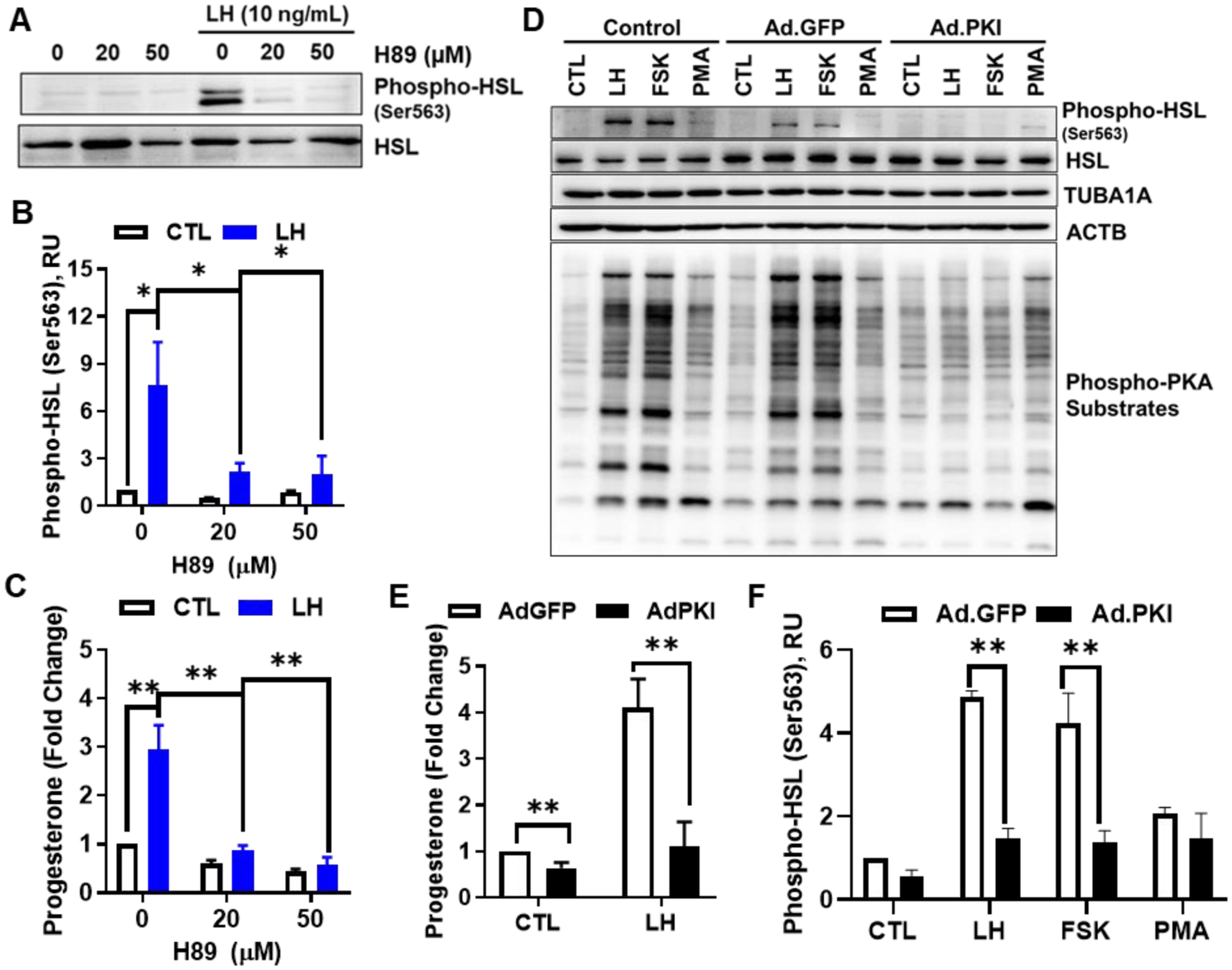
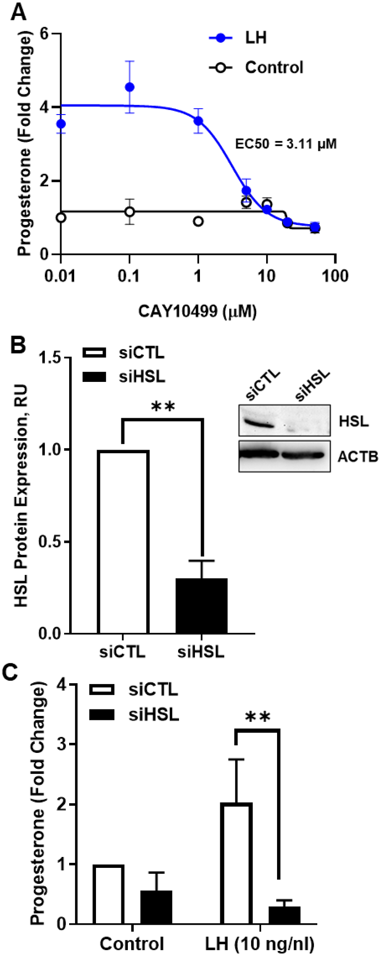
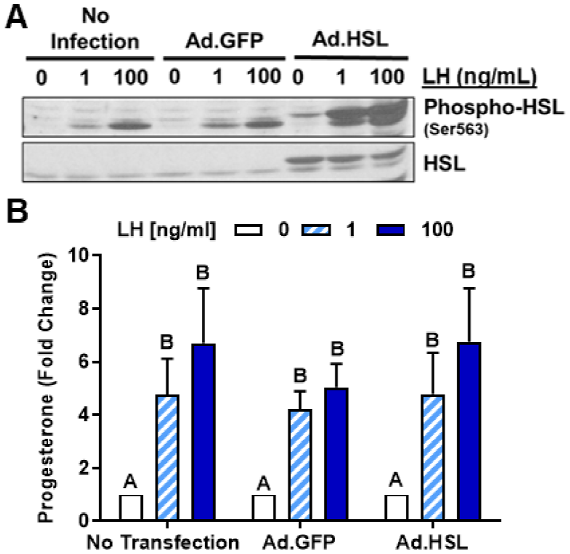
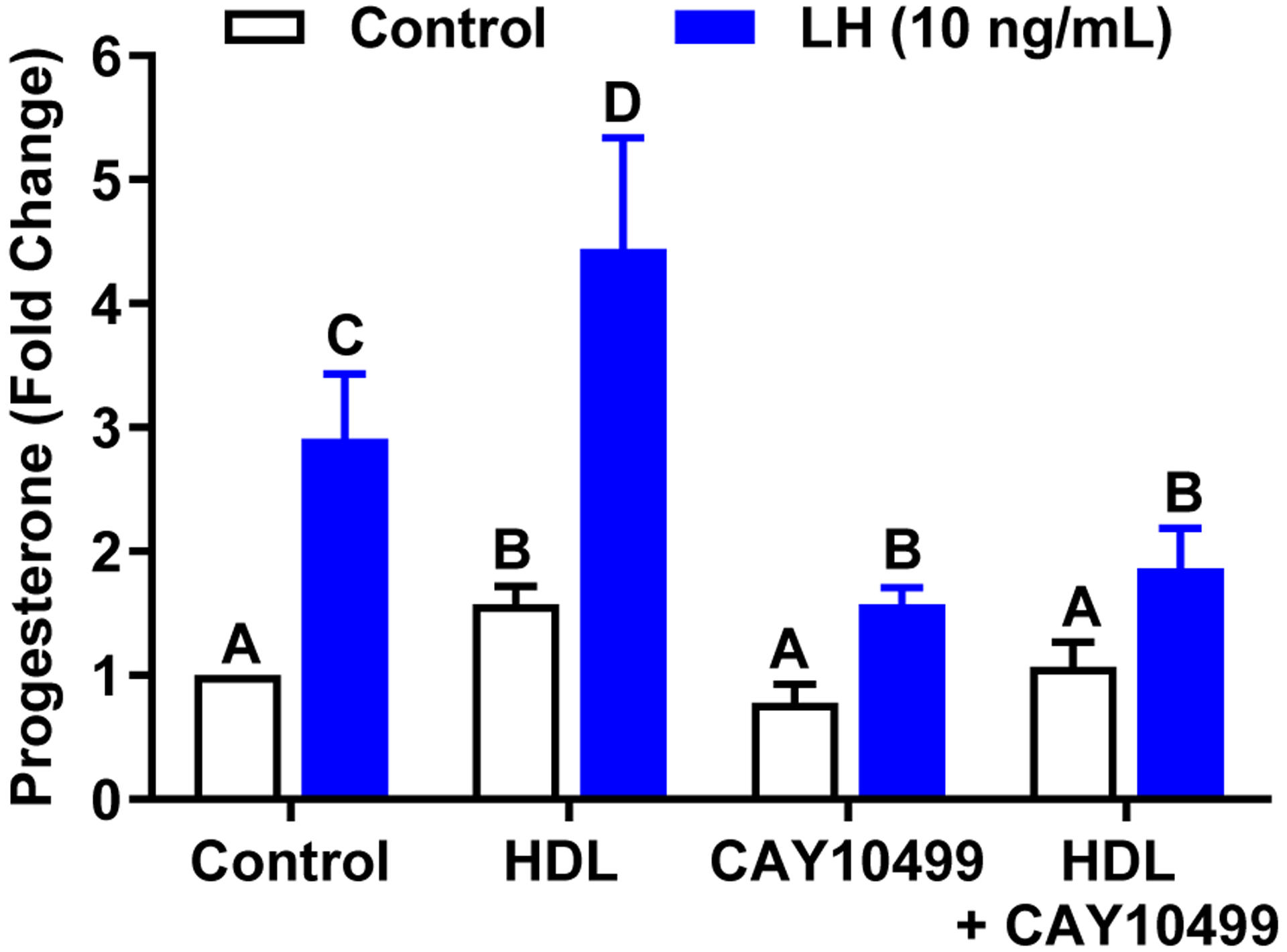
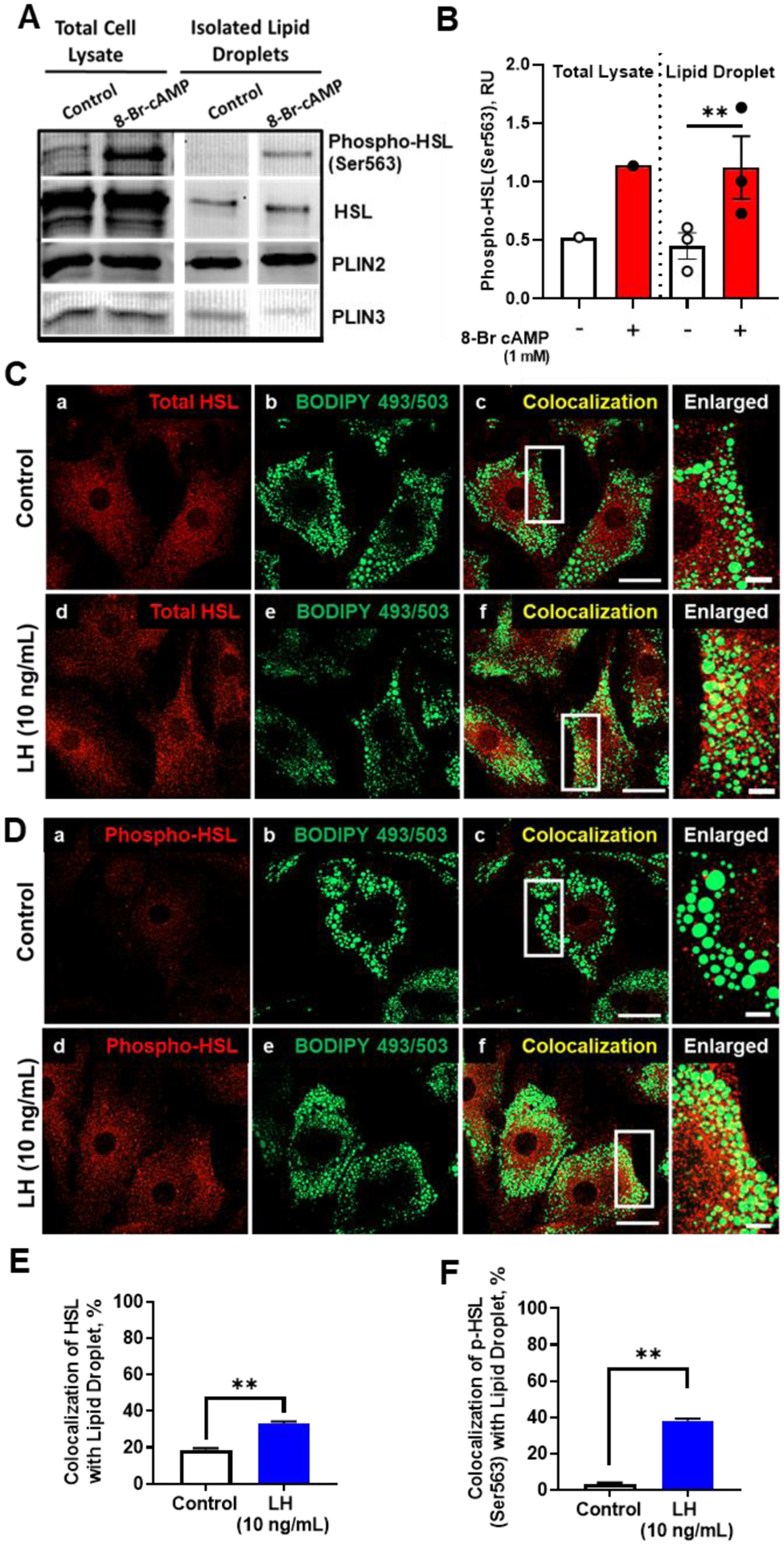
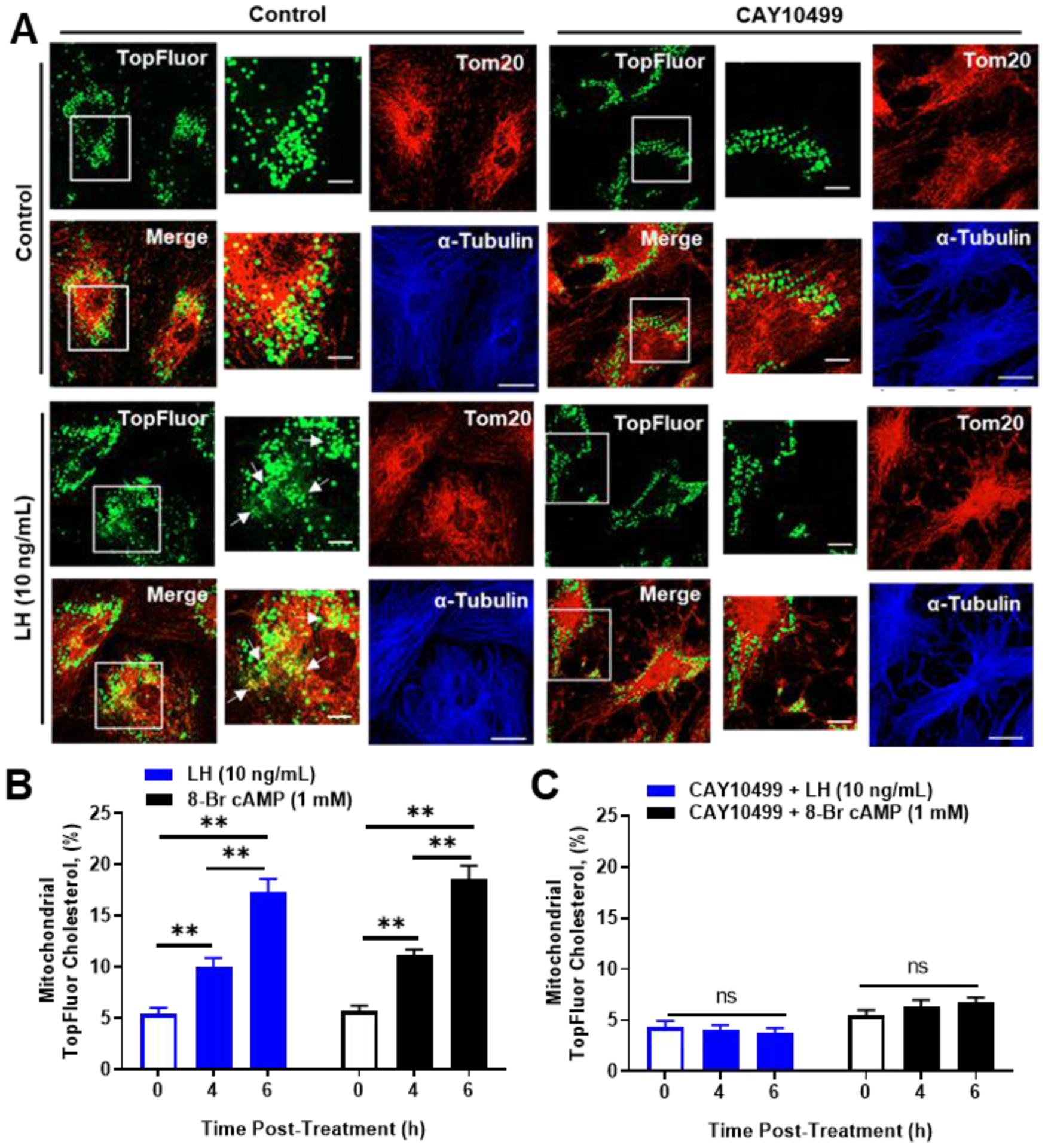
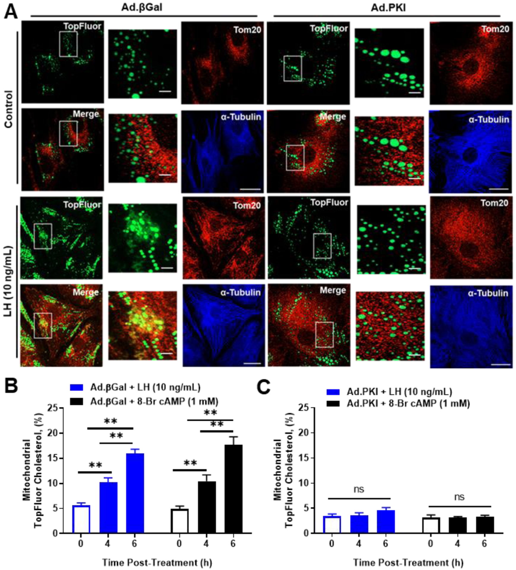
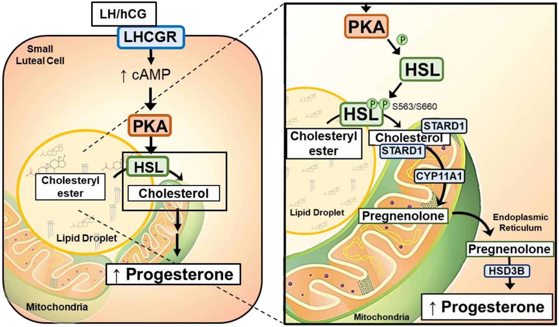
The corpus luteum is a transient endocrine gland that synthesizes and secretes the steroid hormone, progesterone, which is vital for establishment and maintenance of pregnancy. Luteinizing hormone (LH) via activation of protein kinase A (PKA) acutely stimulates luteal progesterone synthesis via a complex process, converting cholesterol via a series of enzymatic reactions, into progesterone. Lipid droplets in steroidogenic luteal cells store cholesterol in the form of cholesterol esters, which are postulated to provide substrate for steroidogenesis. Early enzymatic studies showed that hormone sensitive lipase (HSL) hydrolyzes luteal cholesterol esters. In this study, we tested whether HSL is a critical mediator of the acute actions of LH on luteal progesterone production. Using LH-responsive bovine small luteal cells our results reveal that LH, forskolin, and 8-Br cAMP-induced PKA-dependent phosphorylation of HSL at Ser563 and Ser660, events known to promote HSL activity. Small molecule inhibition of HSL activity and siRNA-mediated knock down of HSL abrogated LH-induced progesterone production. Moreover, western blotting and confocal microscopy revealed that LH stimulates phosphorylation and translocation of HSL to lipid droplets. Furthermore, LH increased trafficking of cholesterol from the lipid droplets to the mitochondria, which was dependent on both PKA and HSL activation. Taken together, these findings identify a PKA/HSL signaling pathway in luteal cells in response to LH and demonstrate the dynamic relationship between PKA, HSL, and lipid droplets in luteal progesterone synthesis.

Keywords: corpus luteum; hormone sensitive lipase; luteinizing hormone; protein kinase A; steroidogenesis.


Figures

Figure 1:
Figure 1:
Expression of Hormone Sensitive Lipase (LIPE/HSL) and components of the steroidogenic machinery in the bovine follicular and luteal cells. Microarray analysis, Western blotting and immunohistochemistry was used to determine the expression of hormone sensitive lipase (LIPE/HSL) in bovine follicular (theca and granulosa) and luteal (small and large) cells. A, Microarray analysis of LIPE/HSL in bovine GC (n = 4; open black bar), TC (n = 3; open blue bar), SLC (n = 3; closed blue bars), and LLC (n = 3; closed black bars). B, Freshly isolated bovine granulosa cell (GC), large luteal cell (LLC), theca cell (TC), small luteal cell (SLC), and mixed luteal cells (mLC) were used to determine the expression of steroidogenic proteins. Representative western blot analysis of steroidogenic protein expression from bovine GC, LLC, TC, SLC, and mLC. Hormone sensitive lipase (HSL); Steroidogenic acute regulatory protein (STAR); Cholesterol side‐chain cleavage enzyme (CYP11A1); 3beta‐Hydroxysteroid dehydrogenase (HSD3B); Beta‐actin (ACTB; loading control). C, Quantification of Western blot analysis of HSL in bovine GC, LLC, TC, SLC, and mixed LC (mean and SEM, n = 3. D, Representative immunohistochemistry micrographs showing expression of HSL in follicles and corpus luteum. Large arrows point to corpus luteum and follicle (a); Negative controls (b and c); HSL staining in GC and TC (d); HSL staining in corpus luteum. Arrows point to endothelial cells. (e). Micron bar represents 5 and 1 μm
Figure 2:
Figure 2:
Effects of luteinizing hormone (LH) on phosphorylation of hormone sensitive lipase (HSL). Small luteal cells were treated for up to 240 minutes with luteinizing hormone (LH; 0‐100 ng/mL), forskolin (10 μM), or 8‐Br cAMP (1 mM) to determine the influence of LH on stimulation of hormone sensitive lipase (HSL). A, Representative western blot analysis for phospho‐ and total‐HSL protein expression in cells treated with increasing concentrations of LH (0‐100 ng/mL) for 30 minutes. B, Quantitative analysis of phospho‐HSL (Ser563) protein expression in cells treated with increasing concentrations of LH. Bars represent mean fold changes (means ± SEM). Bars with different lettersabcd differ significantly within treatment (P < .05). C, Representative western blot analysis for phospho‐ and total‐HSL protein expression in cells treated with LH, forskolin or 8‐Br cAMP. D, Small bovine luteal cells were treated with LH (10 ng/mL) or 8‐Br cAMP (1 mM) for 30 minutes. Representative micrograph showing the effects of LH or 8‐Br cAMP on phosphorylation of HSL (Ser563) in small luteal cells. Control (a‐b), LH (c‐d), and 8‐Br cAMP (e‐f). E, Quantitative analyses of the mean fluorescence intensity of phospho‐HSL (Ser563) in response to treatment with LH or 8‐Br cAMP. Bars represent means ± SEM (n = 3). **Significant difference between treatments as compared to Control, P < .01
Figure 3:
Figure 3:
Effects of Protein Kinase A (PKA) on luteinizing hormone (LH)‐induced HSL phosphorylation and progesterone production in small luteal cells. Panels A‐C: Luteal cells were pretreated with H89, and then, stimulated with LH (10 ng/mL). A, Representative western blot analysis for phospho‐ and total‐HSL protein expression in cells treated with H89 and LH. B, Densitometric analyses of phospho‐HSL (Ser563) protein expression obtained from cells treated with H89 in presence or absence of LH (10 ng/mL). C, Progesterone production by luteal cells pretreated with H89 and stimulated with control or LH for 4 hours. D, Replication‐deficient adenoviruses (Ad) containing the green fluorescent protein (Ad.GFP) and the endogenous inhibitor of PKA (Ad.PKI) were utilized to overexpress GFP or PKI in small luteal cells. After 48 hours, luteal cells were equilibrated and treated for 30 minutes with LH (10 ng/ml), forskolin (FSK, 10 μM), or the protein kinase C activator phorbol 12‐myristate 13‐acetate (PMA; 2 nM). Representative western blot analysis for phospho‐ and total‐HSL protein and total PKA‐substrates. E, Progesterone production by luteal cells transfected with Ad.GFP or Ad.PKI 4 hours posttreatment with control media (CTL) or LH. F, Densitometric analyses of phospho‐HSL (Ser563) protein expression obtained from luteal cells transfected with Ad.GFP (open bars) or Ad.PKI (closed bars) 1‐h posttreatment with LH, FSK, or PMA. Data represented as means ± SEM, n = 3. *Significant difference within treatment as compared to control, P < .05. **, P < .01
Figure 4:
Figure 4:
Effects of Hormone sensitive lipase (HSL) on luteinizing hormone (LH)‐induced progesterone production. A, Luteal cells were pretreated for 1 hour with increasing concentrations of CAY10499 (0 ‐ 100 μM), and then, treated for 4 hours with control (open black line) or LH (closed blue line). Progesterone was measured by ELISA. B, HSL mRNA was silenced using siRNA targeting HSL (siHSL) in small luteal cells. Representative western blot analysis (insert) showing expression of total‐HSL protein expression in siCTL and siHSL knockdown cells. Densitometric analyses of HSL protein expression obtained from siCTL (open bar) and siHSL (closed bar) validating successful knockdown. C, Small luteal cells were transfected with siCTL or siHSL and treated with control media (Control) or LH (10 ng/ml) for 4 hours. Progesterone was measured by ELISA. All data are represented as means ± SEM, n = 3. **Significant difference (P < .01) as compared to control
Figure 5:
Figure 5:
Effects of Hormone Sensitive Lipase (HSL) on luteinizing hormone (LH)‐induced progesterone production in small luteal cells. Replication‐deficient adenovirus (Ad) containing the complete sequence of endogenous green fluorescent protein (GFP) cDNA (Ad.GFP) and Hormone Sensitive Lipase (HSL) cDNA (Ad.HSL) was utilized to overexpress GFP or HSL in luteal cells. A, Representative western blot analysis for phospho‐ and total‐HSL protein expression in cells transfected with Ad. GFP or Ad.HSL in response stimulation with LH (0‐100 ng/mL). B, Progesterone production by enriched small luteal cells transfected with Ad.GFP or Ad.HSL after 4 hours treatment with 0‐100 ng/mL LH. Bars represent mean fold changes (means ± SEM, n = 4). Bars with different lettersabcd differ significantly within treatment (P < .05)
Figure 6:
Figure 6:
Lipoprotein‐mediated progesterone synthesis is prevented by CAY10499. The role of Hormone Sensitive Lipase (HSL) on processing in small luteal cells. To determine if HSL plays a role in HDL processing prior to progesterone biosynthesis, small luteal cells were pretreated with HDL (500 mg/mL), CAY10499 (10 μM), or a combination of HDL and CAY10499. Cells were then stimulated with control medium or LH for 4 hours. Data are represented as means ± SEM, n = 3. Bars with different lettersabcd differ significantly within treatment vs. control (P < .05)
Figure 7:
Figure 7:
Effects of LH on colocalization of phospho‐HSL and HSL with lipid droplets. A, Luteal tissue (punch biopsies) were treated with 8‐Br‐cAMP (1 mM) for 30 minutes and luteal lipid droplets (LDs) were isolated. Representative Western blot of whole tissue lysates and isolated LDs. B, Densitometric analyses of Western blots of phospho‐HSL (Ser563) from lysates and isolated LDs. C, Representative micrographs of HSL (a and d), BODIPY (b and e), colocalization of HSL and BODIPY (c and f), and enlarged image (white box corresponding to adjacent colocalization image) following 30 minutes treatment with control medium or LH. D, Representative micrographs of phospho‐HSL (Ser563) (a and d), BODIPY (b and e), colocalization of phospho‐HSL and BODIPY (c and f), and enlarged image (white box corresponding to adjacent colocalization image) following 30 minutes treatment with LH. E and F, Quantitative analysis of the colocalization of BODIPY with HSL (E) or with phospho‐HSL Ser563 (F) from control and treated cells. Data are represented as means ± SEM, n = 3. **Significant difference, P < .01. Micron bar represents 20 and 2 μm (inset)
Figure 8:
Figure 8:
Effects of Luteinizing hormone (LH) and CAY10499 on trafficking of cholesterol from lipid droplets to mitochondria. Small luteal cells were preloaded with TopFluor Cholesterol for 48 hours. Following cholesterol loading cells were treated with aminoglutethimide (50 μM) and/or CAY10499 (50 μM) for 1‐h prior to stimulation with Control medium, LH (10 ng/mL) or 8‐Br cAMP (1 mM). A, Representative micrographs of (left to right) of Controls: TopFluor Cholesterol, Tom20, merge of TopFluor Cholesterol, and α‐Tubulin from cells treated with Control medium or CAY10499. Representative micrographs of (left to right) of treatment with LH (6 hours): From (left to right) TopFluor Cholesterol, Tom20, merge of TopFluor Cholesterol, and α‐Tubulin from cells treated with LH or LH + CAY10499. Enlarged images correspond to white boxes in the adjacent image. B, Mean accumulation of TopFluor Cholesterol in controls and cells stimulated with LH or 8‐Br cAMP. D, Mean accumulation of TopFluor Cholesterol from cells pretreated with CAY10499, then stimulated with LH or 8‐Br cAMP). Data are represented as means ± SEM, n = 3. **Significant differences between treatments compared to control, P < .01. Micron bar represents 20 μm and 2 μm (inset)
Figure 9:
Figure 9:
Effects of Luteinizing hormone (LH) and PKA on trafficking of cholesterol from lipid droplets to mitochondria. Replication‐deficient adenoviruses (Ad) containing β‐galactosidase cDNA (Ad.βGal) and Protein Kinase Inhibitor cDNA (Ad.PKI) were utilized to overexpress βGal (control) or PKI in luteal cells. Following infection, small luteal cells were preloaded with TopFluor Cholesterol for 48 hours. Following cholesterol loading cells were treated with aminoglutethimide (50 μM) for 1‐h prior to stimulation with LH (10 ng/mL) or 8‐Br cAMP (1 mM). A, Representative micrographs of (left to right) of Controls: TopFluor Cholesterol, Tom20, merge of TopFluor Cholesterol, and α‐Tubulin from cells treated with Ad.βGal or Ad.PKI. Representative micrographs of (left to right) of cells treated with LH (6 hours): From (left to right) TopFluor Cholesterol, Tom20, merge of TopFluor Cholesterol, and α‐Tubulin from cells treated with LH + Ad.βGal or LH + Ad.PKI. Enlarged images correspond to white boxes in the adjacent image. B, Mean mitochondrial accumulation of TopFluor Cholesterol in Ad.βGal transfected cells treated with control, LH or 8‐Br cAMP. C, Mean mitochondrial accumulation of TopFluor Cholesterol in Ad.PKI transfected cells treated with control, LH or 8‐Br cAMP. Data are represented as means ± SEM, n = 3. **Significant difference between treatment as compared to control, P < .01; ns, P > .05; Micron bar represents 20 and 2 μm (inset)
Figure 10:
Figure 10:
Proposed mechanism of action of Luteinizing hormone (LH) in bovine luteal cells. LH activates cAMP/PKA signaling. PKA phosphorylates HSL, which allows it to localize with lipid droplets and enhance its activity. As a result, HSL stimulates the hydrolysis of stores cholesteryl esters releasing cholesterol which is available for transport to the mitochondria by the constitutively expressed transporter steroidogenic acute regulatory protein (STAR). Cholesterol is converted to pregnenolone in the mitochondria, and then, to progesterone by microsomal enzymes in the endoplasmic reticulum (ER)

Similar articles

Cited by

References

    1. Daya S, Ward S, Burrows E J. A. j. o. o., and gynecology. (1988) Progesterone profiles in luteal phase defect cycles and outcome of progesterone treatment in patients with recurrent spontaneous abortion. 158, 225–232 - PubMed
    1. Rueda BR, Hendry IR, Hendry WJ III, Stormshak F, Slayden O, and Davis JS (2000) Decreased progesterone levels and progesterone receptor antagonists promote apoptotic cell death in bovine luteal cells. Biology of reproduction 62, 269–276 - PubMed
    1. Holesh JE, and Lord M (2019) Physiology, ovulation In StatPearls [Internet], StatPearls Publishing - PubMed
    1. O’Shea JD, Rodgers RJ, and D’Occhio MJ (1989) Cellular composition of the cyclic corpus luteum of the cow. J Reprod Fertil 85, 483–487 - PubMed
    1. Hansel W, Alila HW, Dowd JP, and Milvae RA (1991) Differential origin and control mechanisms in small and large bovine luteal cells. J Reprod Fertil Suppl 43, 77–89 - PubMed

Publication types