. 2020 Nov;297(2):E252-E262.
doi: 10.1148/radiol.2020202352. Epub 2020 Jul 2.

Increased Incidence of Barotrauma in Patients with COVID-19 on Invasive Mechanical Ventilation

Affiliations

Increased Incidence of Barotrauma in Patients with COVID-19 on Invasive Mechanical Ventilation

Georgeann McGuinness et al. Radiology. 2020 Nov.

Abstract

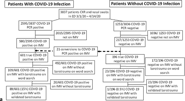
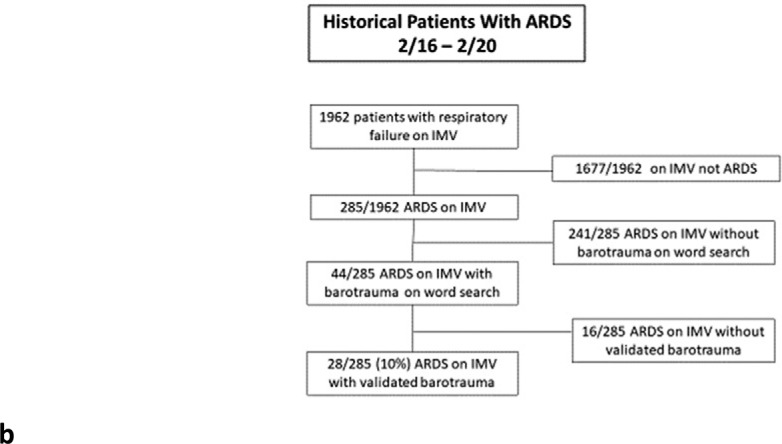
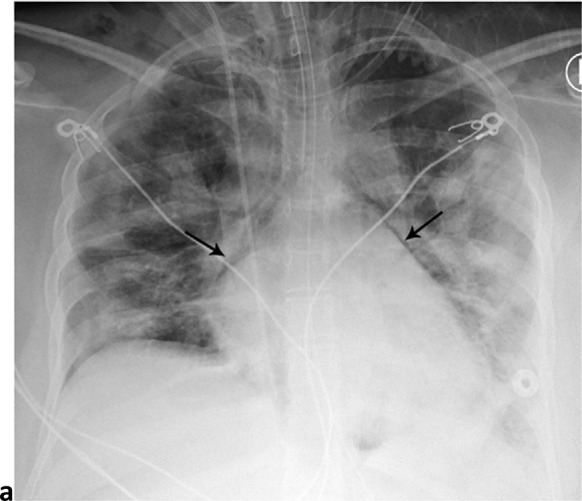
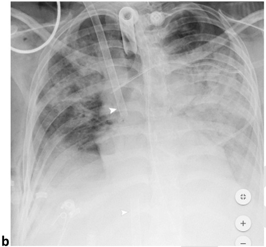
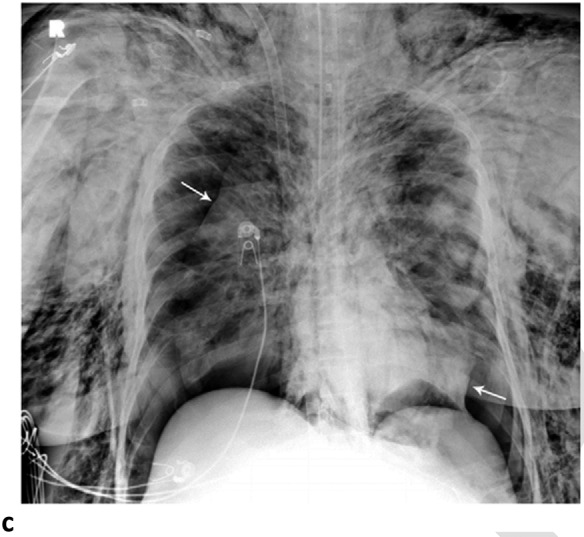
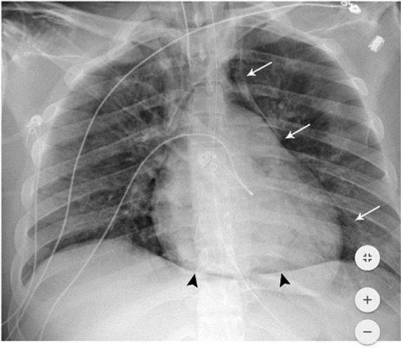
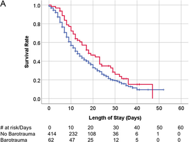
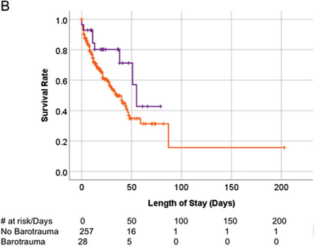
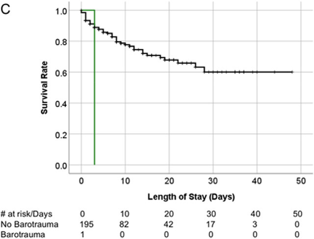
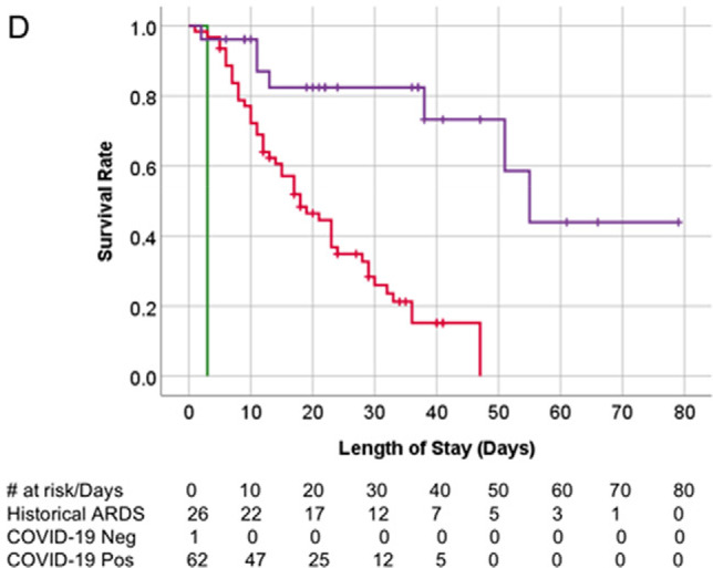
Background A high number of patients with coronavirus disease 2019 (COVID-19) pneumonia who had barotrauma related to invasive mechanical ventilation at the authors' institution were observed. Purpose To determine if the rate of barotrauma in patients with COVID-19 infection was greater than in other patients requiring invasive mechanical ventilation at the authors' institution. Materials and Methods In this retrospective study, clinical and imaging data of patients seen between March 1, 2020, and April 6, 2020, who tested positive for COVID-19 and experienced barotrauma associated with invasive mechanical ventilation, were compared with patients without COVID-19 infection during the same period. Historical comparison was made to barotrauma rates of patients with acute respiratory distress syndrome from February 1, 2016, to February 1, 2020, at the authors' institution. Comparison of patient groups was performed using categoric or continuous statistical testing as appropriate, with multivariable regression analysis. Patient survival was assessed using Kaplan-Meier curves analysis. Results A total of 601 patients with COVID-19 infection underwent invasive mechanical ventilation (mean age, 63 years ± 15 [standard deviation]; 71% men). Of the total, there were 89 (15%) patients with one or more barotrauma events for a total of 145 barotrauma events (24% overall events) (95% confidence interval [CI]: 21%, 28%). During the same period, 196 patients without COVID-19 infection (mean age, 64 years ± 19; 52% men) with invasive mechanical ventilation had one barotrauma event (0.5%; 95% CI: 0%, 3%; P < .001 vs the group with COVID-19 infection). Of 285 patients with acute respiratory distress syndrome on invasive mechanical ventilation during the previous 4 years (mean age, 68 years ± 17; 60% men), 28 patients (10%) had 31 barotrauma events, with an overall barotrauma rate of 11% (95% CI: 8%, 15%; P < .001 vs the group with COVID-19 infection). Barotrauma is an independent risk factor for death in COVID-19 (odds ratio = 2.2; P = .03) and is associated with a longer hospital stay (odds ratio = 0.92; P < .001). Conclusion Patients with coronavirus disease 2019 (COVID-19) infection and invasive mechanical ventilation had a higher rate of barotrauma than patients with acute respiratory distress syndrome and patients without COVID-19 infection. © RSNA, 2020 Online supplemental material is available for this article.


Figures

(a) Patients with and without COVID-19 infection flowchart for inclusion and exclusion. (b) Historical ARDS patient flowchart for inclusion and exclusion. ARDS = Acute Respiratory Distress Syndrome, IMV = Invasive Mechanical Ventilation, PCR = Polymerase Chain Reaction.
Figure 1a:
(a) Patients with and without COVID-19 infection flowchart for inclusion and exclusion. (b) Historical ARDS patient flowchart for inclusion and exclusion. ARDS = Acute Respiratory Distress Syndrome, IMV = Invasive Mechanical Ventilation, PCR = Polymerase Chain Reaction.
Figure 1b:
(a) Patients with and without COVID-19 infection flowchart for inclusion and exclusion. (b) Historical ARDS patient flowchart for inclusion and exclusion. ARDS = Acute Respiratory Distress Syndrome, IMV = Invasive Mechanical Ventilation, PCR = Polymerase Chain Reaction.
Barotrauma events separated by 14 days. 64-year-old man with diabetes and hypertension, intubated 4 days post admission. (a) Frontal chest radiograph demonstrates a large left pneumothorax 6 days after intubation (arrow). (b) He developed a large right pneumothorax 14 days later, 20 days after intubation. There is a left pleural pigtail catheter (black arrow) and re-expansion of the left lung. He underwent tracheostomy 11 days after intubation. Note hazy interstitial densities throughout his lungs.
Figure 2a:
Barotrauma events separated by 14 days. 64-year-old man with diabetes and hypertension, intubated 4 days post admission. (a) Frontal chest radiograph demonstrates a large left pneumothorax 6 days after intubation (arrow). (b) He developed a large right pneumothorax 14 days later, 20 days after intubation. There is a left pleural pigtail catheter (black arrow) and re-expansion of the left lung. He underwent tracheostomy 11 days after intubation. Note hazy interstitial densities throughout his lungs.
Figure 2b:
Barotrauma events separated by 14 days. 64-year-old man with diabetes and hypertension, intubated 4 days post admission. (a) Frontal chest radiograph demonstrates a large left pneumothorax 6 days after intubation (arrow). (b) He developed a large right pneumothorax 14 days later, 20 days after intubation. There is a left pleural pigtail catheter (black arrow) and re-expansion of the left lung. He underwent tracheostomy 11 days after intubation. Note hazy interstitial densities throughout his lungs.
Pneumomediastinum and Bilateral Pneumothoraces, Separated by 7 days. 20-year-old woman intubated 5 days after admission. (a) Frontal chest radiograph depicts moderate pneumomediastinum (black arrows) and subcutaneous emphysema. (b) Frontal radiograph 3 days later demonstrates resolution of pneumomediastinum, and persistent mild subcutaneous emphysema. The superior and inferior extracorporeal membrane oxygenation catheters (arrowheads) were placed the preceding day. (c). Four days later, she developed large bilateral pneumothoraces (white arrows), and extensive subcutaneous emphysema.
Figure 3a:
Pneumomediastinum and Bilateral Pneumothoraces, Separated by 7 days. 20-year-old woman intubated 5 days after admission. (a) Frontal chest radiograph depicts moderate pneumomediastinum (black arrows) and subcutaneous emphysema. (b) Frontal radiograph 3 days later demonstrates resolution of pneumomediastinum, and persistent mild subcutaneous emphysema. The superior and inferior extracorporeal membrane oxygenation catheters (arrowheads) were placed the preceding day. (c). Four days later, she developed large bilateral pneumothoraces (white arrows), and extensive subcutaneous emphysema.
Figure 3b:
Pneumomediastinum and Bilateral Pneumothoraces, Separated by 7 days. 20-year-old woman intubated 5 days after admission. (a) Frontal chest radiograph depicts moderate pneumomediastinum (black arrows) and subcutaneous emphysema. (b) Frontal radiograph 3 days later demonstrates resolution of pneumomediastinum, and persistent mild subcutaneous emphysema. The superior and inferior extracorporeal membrane oxygenation catheters (arrowheads) were placed the preceding day. (c). Four days later, she developed large bilateral pneumothoraces (white arrows), and extensive subcutaneous emphysema.
Figure 3c:
Pneumomediastinum and Bilateral Pneumothoraces, Separated by 7 days. 20-year-old woman intubated 5 days after admission. (a) Frontal chest radiograph depicts moderate pneumomediastinum (black arrows) and subcutaneous emphysema. (b) Frontal radiograph 3 days later demonstrates resolution of pneumomediastinum, and persistent mild subcutaneous emphysema. The superior and inferior extracorporeal membrane oxygenation catheters (arrowheads) were placed the preceding day. (c). Four days later, she developed large bilateral pneumothoraces (white arrows), and extensive subcutaneous emphysema.
Pneumomediastinum and pneumopericardium. 18-year-old man without significant medical history was intubated 5 days after admission, and developed pneumomediastinum and pneumopericardium the same day. Frontal chest radiograph depicts mediastinal air bilaterally (white arrows). The ‘continuous diaphragm sign’ (black arrowheads) indicates air beneath the heart. Note diffuse hazy interstitial lung markings.
Figure 4:
Pneumomediastinum and pneumopericardium. 18-year-old man without significant medical history was intubated 5 days after admission, and developed pneumomediastinum and pneumopericardium the same day. Frontal chest radiograph depicts mediastinal air bilaterally (white arrows). The ‘continuous diaphragm sign’ (black arrowheads) indicates air beneath the heart. Note diffuse hazy interstitial lung markings.
Kaplan-Meier survival curves. (A) Overall survival of patients with COVID-19 infection on invasive mechanical ventilation. Patients with and without barotrauma represented by red and blue curves respectively, with no difference in overall survival (p>.05). (B) Overall survival of historical ARDS invasive mechanically ventilated patients. Patients with and without barotrauma represented by purple and orange curves respectively, with no difference in overall survival (p>.05). (C) Overall survival of patients without COVID-19 infection on invasive mechanical ventilation. Patients with and without barotrauma represented by green and black curves respectively, with longer survival for patients without barotrauma (p=.01). (D) Overall survival of invasive mechanically ventilated patients with barotrauma. Patients with ARDS, with and without COVID-19 infection represented by purple, red and green curves respectively, showed longer survival for patients in the ARDS group (p<.001).
Figure 5a:
Kaplan-Meier survival curves. (A) Overall survival of patients with COVID-19 infection on invasive mechanical ventilation. Patients with and without barotrauma represented by red and blue curves respectively, with no difference in overall survival (p>.05). (B) Overall survival of historical ARDS invasive mechanically ventilated patients. Patients with and without barotrauma represented by purple and orange curves respectively, with no difference in overall survival (p>.05). (C) Overall survival of patients without COVID-19 infection on invasive mechanical ventilation. Patients with and without barotrauma represented by green and black curves respectively, with longer survival for patients without barotrauma (p=.01). (D) Overall survival of invasive mechanically ventilated patients with barotrauma. Patients with ARDS, with and without COVID-19 infection represented by purple, red and green curves respectively, showed longer survival for patients in the ARDS group (p<.001).
Figure 5b:
Kaplan-Meier survival curves. (A) Overall survival of patients with COVID-19 infection on invasive mechanical ventilation. Patients with and without barotrauma represented by red and blue curves respectively, with no difference in overall survival (p>.05). (B) Overall survival of historical ARDS invasive mechanically ventilated patients. Patients with and without barotrauma represented by purple and orange curves respectively, with no difference in overall survival (p>.05). (C) Overall survival of patients without COVID-19 infection on invasive mechanical ventilation. Patients with and without barotrauma represented by green and black curves respectively, with longer survival for patients without barotrauma (p=.01). (D) Overall survival of invasive mechanically ventilated patients with barotrauma. Patients with ARDS, with and without COVID-19 infection represented by purple, red and green curves respectively, showed longer survival for patients in the ARDS group (p<.001).
Figure 5c:
Kaplan-Meier survival curves. (A) Overall survival of patients with COVID-19 infection on invasive mechanical ventilation. Patients with and without barotrauma represented by red and blue curves respectively, with no difference in overall survival (p>.05). (B) Overall survival of historical ARDS invasive mechanically ventilated patients. Patients with and without barotrauma represented by purple and orange curves respectively, with no difference in overall survival (p>.05). (C) Overall survival of patients without COVID-19 infection on invasive mechanical ventilation. Patients with and without barotrauma represented by green and black curves respectively, with longer survival for patients without barotrauma (p=.01). (D) Overall survival of invasive mechanically ventilated patients with barotrauma. Patients with ARDS, with and without COVID-19 infection represented by purple, red and green curves respectively, showed longer survival for patients in the ARDS group (p<.001).
Figure 5d:
Kaplan-Meier survival curves. (A) Overall survival of patients with COVID-19 infection on invasive mechanical ventilation. Patients with and without barotrauma represented by red and blue curves respectively, with no difference in overall survival (p>.05). (B) Overall survival of historical ARDS invasive mechanically ventilated patients. Patients with and without barotrauma represented by purple and orange curves respectively, with no difference in overall survival (p>.05). (C) Overall survival of patients without COVID-19 infection on invasive mechanical ventilation. Patients with and without barotrauma represented by green and black curves respectively, with longer survival for patients without barotrauma (p=.01). (D) Overall survival of invasive mechanically ventilated patients with barotrauma. Patients with ARDS, with and without COVID-19 infection represented by purple, red and green curves respectively, showed longer survival for patients in the ARDS group (p<.001).

References

    1. Petrilli CM, Jones SA, Yang J, et al. . Factors associated with hospitalization and critical illness among 4,103 patients with COVID-19 disease in New York City. medRxiv. 2020:2020.2004.2008.20057794. Accepted for publication, British Journal of Medicine 2020
    1. Anzueto A, Frutos–Vivar F, Esteban A, et al. . Incidence, risk factors and outcome of barotrauma in mechanically ventilated patients. Intensive care medicine. 2004;30(4):612-619. - PubMed
    1. Boussarsar M, Thierry G, Jaber S, Roudot-Thoraval F, Lemaire F, Brochard L. Relationship between ventilatory settings and barotrauma in the acute respiratory distress syndrome. Intensive care medicine. 2002;28(4):406-413. - PubMed
    1. Briel M, Meade M, Mercat A, et al. . Higher vs lower positive end-expiratory pressure in patients with acute lung injury and acute respiratory distress syndrome: systematic review and meta-analysis. Jama. 2010;303(9):865-873. - PubMed
    1. Grasso S, Stripoli T, De Michele M, et al. . ARDSnet ventilatory protocol and alveolar hyperinflation: role of positive end-expiratory pressure. American journal of respiratory and critical care medicine. 2007;176(8):761-767. - PubMed

MeSH terms