Review
. 2020 Aug 3;219(8):e202003131.
doi: 10.1083/jcb.202003131.

Phospholipid ebb and flow makes mitochondria go

Affiliations
Review

Phospholipid ebb and flow makes mitochondria go

Michelle Grace Acoba et al. J Cell Biol. .

Abstract

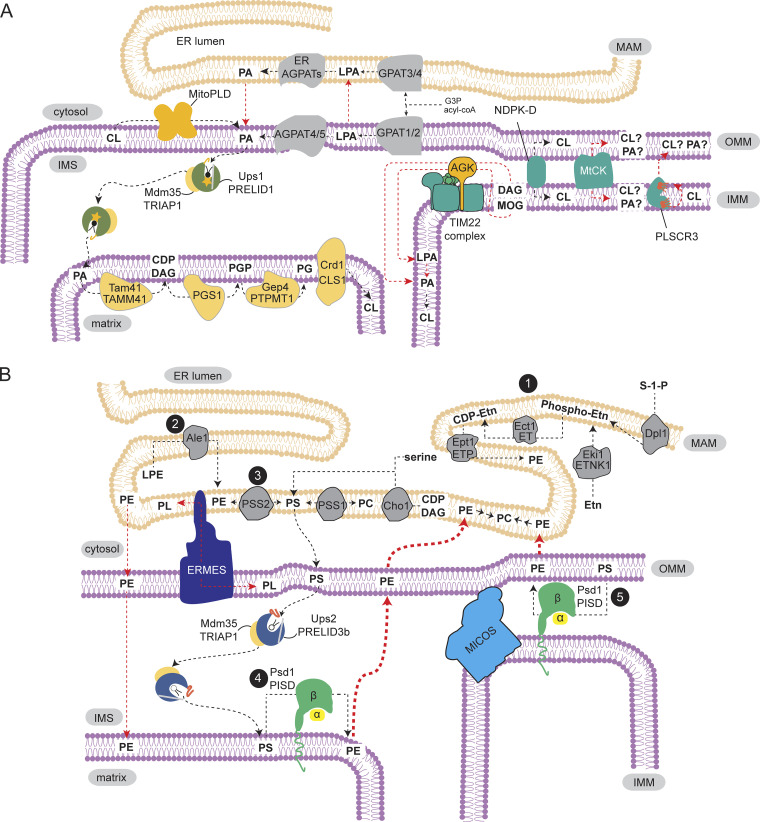
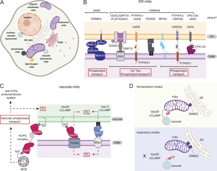
Mitochondria, so much more than just being energy factories, also have the capacity to synthesize macromolecules including phospholipids, particularly cardiolipin (CL) and phosphatidylethanolamine (PE). Phospholipids are vital constituents of mitochondrial membranes, impacting the plethora of functions performed by this organelle. Hence, the orchestrated movement of phospholipids to and from the mitochondrion is essential for cellular integrity. In this review, we capture recent advances in the field of mitochondrial phospholipid biosynthesis and trafficking, highlighting the significance of interorganellar communication, intramitochondrial contact sites, and lipid transfer proteins in maintaining membrane homeostasis. We then discuss the physiological functions of CL and PE, specifically how they associate with protein complexes in mitochondrial membranes to support bioenergetics and maintain mitochondrial architecture.


Figures

Figure 1.
Figure 1.
Biosynthesis of CL and PE. (A) CL production starts with PA, which can be sourced from multiple pathways (Box 1). PA needs to be trafficked to the matrix side of the IMM, where CL biosynthetic enzymes reside. PA shuttling from the OMM to the IMM is performed by Ups1-Mdm35/PRELID1-TRIAP1. Trafficking of CL or PA between OMM and IMM may also involve proteins at contact sites such as NDPK-D and MtCK. PLSCR3 promotes CL externalization to the OMM. It may transport CL or PA from the IMM to OMM or facilitate CL flip-flop from the inner to outer leaflet of the IMM. (B) PE is produced in the ER and mitochondria. ER pathways include the CDP–ethanolamine/Kennedy pathway (1), lysophosphatidylethanolamine acylation in yeast (2), and head group base exchange with PS in mammals (3). In mammals, PS can also be formed via base exchange with PC by PSS2, while in yeast, it is generated from CDP-DAG and serine by Cho1. PS produced in the MAM by PSS1/PSS2/Cho1 is then translocated to the OMM with the help of ER-mitochondrion MCS, such as ERMES in yeast. Ups2-Mdm35/PRELID3b-TRIAP1 transports PS to the IMM, where Psd1/PISD decarboxylates it to PE (4). Tethering of IMM and OMM by MICOS may allow Psd1/PISD to convert PS to PE in the OMM (5). PE made in the mitochondrion is exported to the ER, where it serves as a substrate for PC production. PE from the ER or OMM can also reach the IMM to a limited extent. Unresolved mechanisms are depicted by red arrows. MOG, monoacylglycerol.
Figure 2.
Figure 2.
Mitochondrial membrane contact sites. (A) The mitochondrion establishes contacts with multiple organelles, which are important for various cellular processes, including phospholipid transport. (B) Physical interaction between ER and mitochondria is mediated by the ERMES complex in yeast, whereas a number of ER–mitochondria tethers have been documented in metazoans. SMP lipid-binding domains are present in ERMES, indicating a role in phospholipid shuttling aside from maintaining a close apposition of ER and OMM. ORP5/8 and PDZD8 have ORD and SMP domains, respectively; PDZD8 has not been shown to directly bind/transfer phospholipids. A domain in yeast Vps13 has been shown to possess lipid transport capacity. This domain is conserved in VPS13A and other VPS13 human isoforms. (C) Two types of vCLAMP, Vps39/Ypt7/Tom40 and Vps13/Mcp1 (vacuole counterpart unknown), connect vacuole and mitochondrion, allowing phospholipid (PL) exchange. As a component of the HOPS complex, Vps39 is also relevant for endolysosomal trafficking. Vps39 may facilitate vesicular phospholipid transport to the vacuole or to other parts of the endomembrane system, which can in turn supply phospholipids to the mitochondrion. (D) Metabolic conditions can regulate mitochondrial contact sites. Respiratory conditions lead to Vps39 phosphorylation that prevents vCLAMP formation, favoring OMM contacts with ERMES. Stars, lipid-binding domains; red arrow, unresolved mechanism. A and D were created with Biorender.
Figure 3.
Figure 3.
Phospholipid transport across the IMS. Ups/PRELI family contributes to PA and PS transport between the OMM and IMM. Once lipid is extracted from the OMM and incorporated into the pocket of the transfer protein, Ups/PRELI associates with Mdm35/TRIAP1 and closes its lid, enabling efficient transport. Upon reaching the IMM, the protein complex dissociates, facilitating lipid release. Ups/PRELI proteins are then subjected to proteolytic degradation. (A) A positively charged residue (K58) at the bottom of Ups1/PRELID1’s cavity interacts with the PA head group. PA is extracted from OMM in a process that is perhaps facilitated by the cone-shaped structure of PA that prevents tight packing. The high CL concentration in the IMM helps recruit Ups1/PRELID1. PA transport is stimulated and repressed by high PA and high CL levels, respectively. (B) PS transport by Ups2-Mdm35/PRELID3b-TRIAP1 permits PE production in the IMM. The greater hydrophobicity of the Ω loop and the α3 helix of PRELID3b aids in PS extraction from OMM, even though its cylindrical structure does not support its extraction. High CL amounts promote PS translocation.
Figure 4.
Figure 4.
Physiological functions of mitochondrial phospholipids. (A) Association of phospholipids and proteins in the IMM impacts stability of both protein complexes and membrane phospholipids. Loss of CL leads to RSC disassembly, while loss of OXPHOS proteins (thus disrupting RSC formation) results in faster CL turnover that is thought to elevate the MLCL/CL ratio. The amount of saturated CL also increases in the absence of OXPHOS complexes that induce CL remodeling. (B) CL-supported cristae structure via MICOS integrity. The mammalian MICOS complex is depicted. In the presence of CL, the MIC10 subcomplex, composed of MIC10, QIL1, MIC27, and MIC26, assembles and organizes cristae structure. MIC27 specifically interacts with CL. In the absence of CL, the MIC10 subcomplex is diffused, disrupting cristae morphology. MIC60 subcomplex assembly is independent of CL. (C) Phospholipid involvement in mitochondrial dynamics. (Top) DRP1 modulates membrane restriction in response to the local concentrations of CL and PA. DRP1 is recruited to the OMM and oligomerizes in a CL-dependent manner, leading to membrane constriction. It directly interacts with mitoPLD and binds mitoPLD-produced PA, which in turn suppresses division. (Bottom) MFN and MIGA promote OMM fusion where PA locally accumulates, as a consequence of MitoPLD-mediated CL hydrolysis. MIGA directly interacts with mitoPLD. OPA1 forms a dimer and induces IMM fusion at the CL-enriched sites.

Similar articles

Cited by

References

    1. Aaltonen M.J., Friedman J.R., Osman C., Salin B., di Rago J.P., Nunnari J., Langer T., and Tatsuta T.. 2016. MICOS and phospholipid transfer by Ups2-Mdm35 organize membrane lipid synthesis in mitochondria. J. Cell Biol. 213:525–534. 10.1083/jcb.201602007 - DOI - PMC - PubMed
    1. Acehan D., Vaz F., Houtkooper R.H., James J., Moore V., Tokunaga C., Kulik W., Wansapura J., Toth M.J., Strauss A., et al. . 2011. Cardiac and skeletal muscle defects in a mouse model of human Barth syndrome. J. Biol. Chem. 286:899–908. 10.1074/jbc.M110.171439 - DOI - PMC - PubMed
    1. Achleitner G., Zweytick D., Trotter P.J., Voelker D.R., and Daum G.. 1995. Synthesis and intracellular transport of aminoglycerophospholipids in permeabilized cells of the yeast, Saccharomyces cerevisiae. J. Biol. Chem. 270:29836–29842. 10.1074/jbc.270.50.29836 - DOI - PubMed
    1. Acín-Pérez R., Fernández-Silva P., Peleato M.L., Pérez-Martos A., and Enriquez J.A.. 2008. Respiratory active mitochondrial supercomplexes. Mol. Cell. 32:529–539. 10.1016/j.molcel.2008.10.021 - DOI - PubMed
    1. Adachi Y., Itoh K., Yamada T., Cerveny K.L., Suzuki T.L., Macdonald P., Frohman M.A., Ramachandran R., Iijima M., and Sesaki H.. 2016. Coincident Phosphatidic Acid Interaction Restrains Drp1 in Mitochondrial Division. Mol. Cell. 63:1034–1043. 10.1016/j.molcel.2016.08.013 - DOI - PMC - PubMed

Publication types