. 2020 Jul 2;16(7):e1008601.
doi: 10.1371/journal.ppat.1008601. eCollection 2020 Jul.

Zika virus tropism during early infection of the testicular interstitium and its role in viral pathogenesis in the testes

Affiliations

Zika virus tropism during early infection of the testicular interstitium and its role in viral pathogenesis in the testes

Konstantin A Tsetsarkin et al. PLoS Pathog. .

Abstract

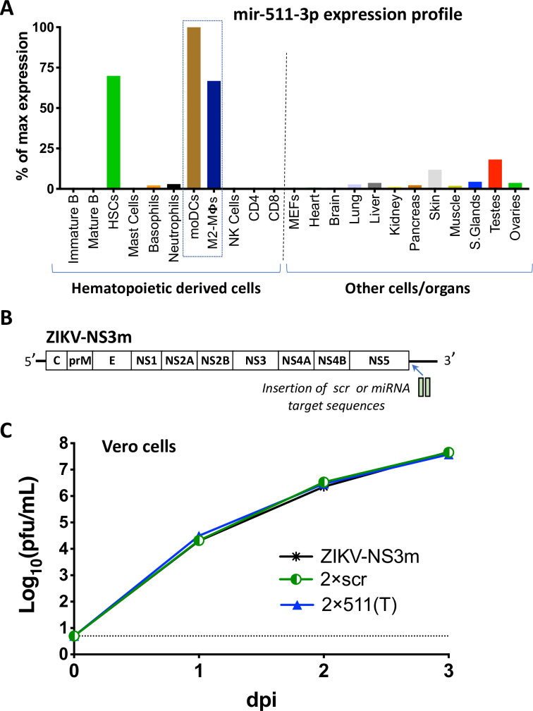
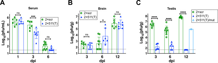
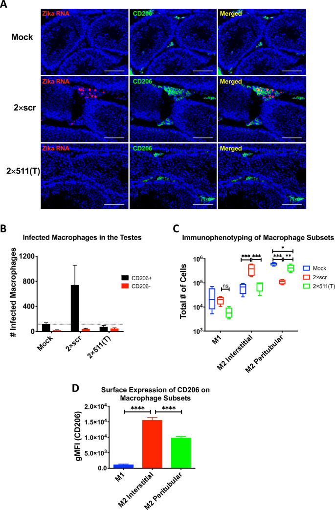
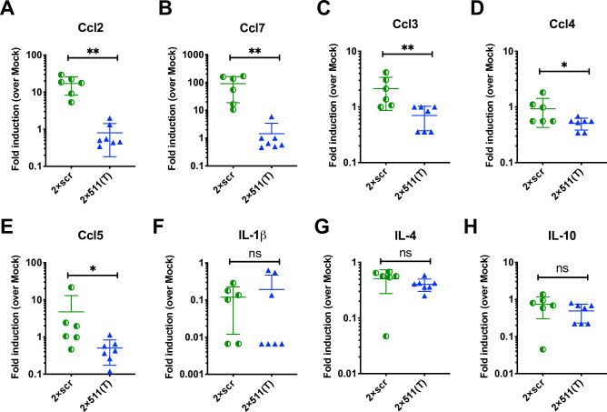
Sexual transmission and persistence of Zika virus (ZIKV) in the testes pose new challenges for controlling virus outbreaks and developing live-attenuated vaccines. It has been shown that testicular infection of ZIKV is initiated in the testicular interstitium, followed by spread of the virus in the seminiferous tubules. This leads to testicular damage and/or viral dissemination into the epididymis and eventually into semen. However, it remains unknown which cell types are targeted by ZIKV in the testicular interstitium, and what is the specific order of infectious events leading to ZIKV invasion of the seminiferous tubules. Here, we demonstrate that interstitial leukocytes expressing mir-511-3p microRNA are the initial targets of ZIKV in the testes, and infection of mir-511-3p-expressing cells in the testicular interstitium is necessary for downstream infection of the seminiferous tubules. Mir-511-3p is expressed concurrently with CD206, a marker of lineage 2 (M2) macrophages and monocyte derived dendritic cells (moDCs). Selective restriction of ZIKV infection of CD206-expressing M2 macrophages/moDCs results in the attenuation of macrophage-associated inflammatory responses in vivo and prevents the disruption of the Sertoli cell barrier in vitro. Finally, we show that targeting of viral genome for mir-511-3p significantly attenuates early ZIKV replication not only in the testes, but also in many peripheral organs, including spleen, epididymis, and pancreas. This incriminates M2 macrophages/moDCs as important targets for visceral ZIKV replication following hematogenous dissemination of the virus from the site of infection.


Conflict of interest statement

The authors have declared that no competing interests exist.

Figures

Fig 1
Fig 1. Characterization of a ZIKV clone containing target sites for M2 macrophage/moDC-specific miRNA.
(A) Expression profile of the mir-511-3p in selected cells and organs of mice. The graph was constructed based on deep sequencing data of the mouse miRNAs previously reported [38]. The expression profile is presented as a proportion of the number of reads for mir-511-3p in the cells/organs to the number of reads for this miRNA in the moDCs, which has the highest expression level among all types of cells or organs. HSCs—hematopoietic stem cells, MΦs-macrophages, NK—natural killer, MEFs–mouse embryonic fibroblasts. (B) Schematic representation of the insertion of scr or mir-511-3p target sequences into the genome of ZIKV-NS3m virus [18]. (C) Growth of ZIKV-NS3m, 2×scr, and 2×511(T) viruses in Vero cells after plasmid DNA transfection. Data show mean virus titer ± standard deviation (SD) in cell culture supernatants, which were collected daily from duplicate flasks of transfected Vero cells. The dashed line indicates the limit of virus detection [0.7 log10(pfu/mL)].
Fig 2
Fig 2. Effect of mir-511-3p target insertions on ZIKV replication in the serum, brain and testes of AG129 mice.
Adult AG129 male mice (n = 6–9 per group) were infected IP with 106 pfu of ZIKV clones containing target sites for mir-511-3p [2×511(T)] or scrambled target sites [2×scr]. Mice were bled and/or sacrificed at the indicated days post infection. Mean viral titer ± SD in the serum (A), brain (B), or testes (C) was determined by titration in Vero cells. The dashed lines indicate the limit of virus detection: 1.5 log10 pfu/mL of serum (A) and 1.7 log10 pfu/g of brain or testis (B-C). Differences between the virus titers were compared using two-way ANOVA (**** p < 0.0001; *** p< 0.001; * p<0.05; ns—denotes not significant, p> 0.05). Except 6 dpi, all data for the 2×scr virus are retrospective [17] and provided here for comparison. Data for 6 dpi for the 2×scr virus was combined from the data reported earlier (n = 6) [17] and newly generated results (n = 3). Viral RNA was isolated from the serum at 1 dpi, brain at 12 dpi and testes at 12 dpi, and the region containing miRNA targets was sequenced. If mutation/deletion was detected in the miRNA targeted region, viral titer for these samples was reported under the name 2×511(T)/mut.
Fig 3
Fig 3. Targeting of ZIKV genome for mir-511-3p prevents infection of CD206 expressing macrophages in the testicular interstitium.
Adult AG129 male mice were mock-inoculated or infected IP with 106 pfu of 2×scr or 2×511(T) virus. For panel (A), mock (n = 1), 2×scr (n = 3) and 2×511(T) (n = 3); for panels (B-D), mock (n = 3), 2×scr (n = 3) and 2×511(T) (n = 4). Mice were sacrificed at 3 dpi and testes were harvested. (A) Testes sections stained for ZIKV RNA (red) by in situ hybridization and CD206 (green) by immunofluorescence co-staining. Scale bars represent 100μm. To identify the ZIKV-infected cells within the CD206-positive and CD206-negative populations, cells from testes at 3 dpi were dissociated into a single cell suspension, fixed, and stained for ZIKV using anti-E protein antibody, 4G2, to detect ZIKV and CD206 (B). Single cell suspensions from testes were stained with CD45, F4/80, CD11c, CD11b, MHC II and CD206 to differentiate various myeloid cell types (C). (D) Expression of CD206 was determined in each cell population identified in C. Statistical significance was determined for (C) and (D) by two way ANOVA and one way ANOVA for multiple comparisons. *, **, ***, **** indicate p<0.05, p<0.01, p≤0.001 and p≤0.0001, respectively.
Fig 4
Fig 4. Infection of testicular macrophages is necessary for the production of macrophage-associated inflammatory mediators.
Testis homogenates from AG129 mice infected with either 2×scr (n = 6) or 2×511(T) (n = 7) or mock inoculated mice (n = 3) were collected at 3dpi and analyzed for chemokine and cytokine production by multiplexed ELISA. Fold induction over values found in mock-inoculated mice is shown as pg/g of tissue. (A-E) macrophage-associated chemokines and (F-H) representative markers of inflammation. Significance was determined by a two-tailed Mann-Whitney test. * indicates p<0.05, ** indicates p<0.01.
Fig 5
Fig 5. Soluble factors produced in the presence of ZIKV-infected macrophages can disrupt the Sertoli cell barrier.
A murine Sertoli cell line, 15-P1, was grown to confluency in transwells. Cells were then exposed to testis homogenates from AG129 mice that were infected with 2×scr (n = 2 for experiment depicted in (A), and n = 3 for experiment depicted in panel (B)), 2×511(T) (n = 3), or mock inoculated (n = 3). One hundred nanograms of murine TNF⍺ was also included as a positive control (A). A voltage was applied across the transwell 24 hours post exposure, and resistance was measured. Transepithelial electrical resistance (TEER) was calculated as ohms/cm2. Since 15-P1 monolayers had grown to different peak TEER values in two independent experiments, raw TEER values are reported separately (A) and (B) for two independent, double blinded experiments. Significance was determined by one way ANOVA. ** indicates p<0.01.
Fig 6
Fig 6. Insertion of multiple copies of mir-511-3p target sequences into distant regions of the ZIKV genome inhibits viral replication in peripheral mouse organs.
(A) Schematic representation of viruses used in the mouse studies. (B-K) Male AG129 mice (5 per group) were infected IP with 106 pfu of C/3’NCR-511(T) or C/3’NCR-scr virus and were bled at 1 dpi and sacrificed at 3 dpi, or were not bled and sacrificed at 12 dpi. (B) Mean virus titer ± SD in the serum at 1 and 3 dpi. (C-D) Mean virus titer ± SD in the testes (C) and epididymis (D) at 3 dpi and 12 dpi. (E-K) Mean virus titer ± SD in the spleen (E), pancreas (F), lung (G), femoris muscle (H), submandibular gland (I) and small intestine (J) at 3 dpi, and in the brain (K) at 12 dpi. The dashed lines indicate the limit of virus detection: in the serum [1.5 log10 pfu/mL]; in the testes, spleen, lung, femoris muscle, submandibular gland and brain [1.7 log10 pfu/g of tissue]; in the pancreas and small intestine [2.7 log10 pfu/g of tissue]; and in the epididymis [0.7 log10 pfu/mouse]. Differences between viral titers in the serum, testes and epididymis were compared using two-way ANOVA, and differences between viral titers in other organs were compared using two-tailed Mann-Whitney test. *** p<0.001; **** p< 0.0001; ns denotes p>0.05.
Fig 7
Fig 7. Immunogenicity and protective efficacy of the ZIKV containing multiple copies of mir-511-3p target sequences in AG129 mice.
(A) Adult male or female AG129 mice were mock inoculated (n = 5) or IP infected with 105 (male) or 103 (female) pfu of C/3’NCR-511(T) or C/3’NCR-scr. On dpi 28 and 56, animals were bled to determine NA titer, which is presented as mean ± SD (B). On dpi 29, animals were challenged IP with 105pfu of wt ZIKV (strain Paraiba_01/2015). At 2 days post challenge (dpc) mice were bled to determine viremia (C, D) and monitored for survival (E, F). The dashed lines indicate the limit of virus detection in the serum [1.5 log10 pfu/mL (C, D)] or indicate the limit of NA titer detection (B). Differences between viral titers in the serum were compared using two-way ANOVA (C, D). Differences between NA titers were compared using two-tailed Mann-Whitney test (B). ** p<0.01; **** p< 0.0001; ns denotes p>0.05. Data for male and female mice is presented as a summary of two and one independent experiments, respectively.

References

    1. Agumadu VC, Ramphul K. Zika Virus: A Review of Literature. Cureus. 2018;10(7):e3025 Epub 2018/09/27. 10.7759/cureus.3025 - DOI - PMC - PubMed
    1. Honein MA, Dawson AL, Petersen EE, Jones AM, Lee EH, Yazdy MM, et al. Birth Defects Among Fetuses and Infants of US Women With Evidence of Possible Zika Virus Infection During Pregnancy. JAMA. 2017;317(1):59–68. Epub 2016/12/14. 10.1001/jama.2016.19006 . - DOI - PubMed
    1. CDC.gov. Data and Statistics on Zika and Pregnancy 2019 [updated March 29, 2019; cited 2019 09/26]. Available from: https://www.cdc.gov/pregnancy/zika/data/index.html.
    1. Miner JJ, Diamond MS. Zika Virus Pathogenesis and Tissue Tropism. Cell Host Microbe. 2017;21(2):134–42. Epub 2017/02/10. 10.1016/j.chom.2017.01.004 - DOI - PMC - PubMed
    1. Mead PS, Hills SL, Brooks JT. Zika virus as a sexually transmitted pathogen. Curr Opin Infect Dis. 2018;31(1):39–44. 10.1097/QCO.0000000000000414 WOS:000424041200006. - DOI - PubMed

Publication types

LinkOut - more resources