Review
. 2021 Feb;162(2):123-134.
doi: 10.1111/imm.13231. Epub 2020 Jul 27.

Schistosome and intestinal helminth modulation of macrophage immunometabolism

Affiliations
Review

Schistosome and intestinal helminth modulation of macrophage immunometabolism

Diana Cortes-Selva et al. Immunology. 2021 Feb.

Abstract

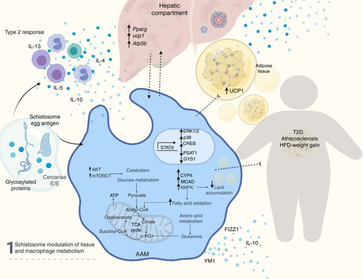
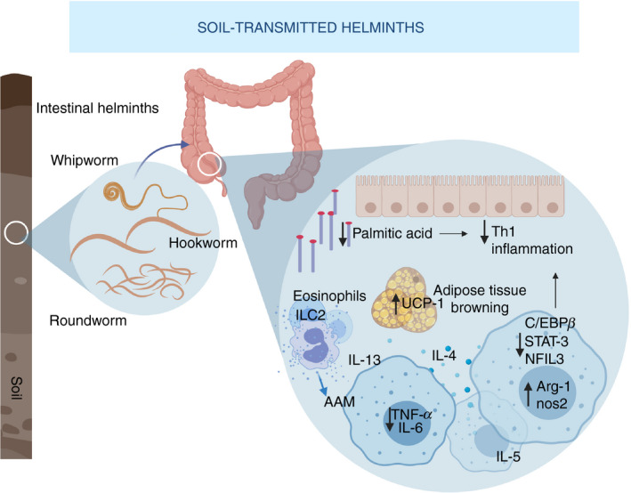
Macrophages are fundamental to sustain physiological equilibrium and to regulate the pathogenesis of parasitic and metabolic processes. The functional heterogeneity and immune responses of macrophages are shaped by cellular metabolism in response to the host's intrinsic factors, environmental cues and other stimuli during disease. Parasite infections induce a complex cascade of cytokines and metabolites that profoundly remodel the metabolic status of macrophages. In particular, helminths polarize macrophages to an M2 state and induce a metabolic shift towards reliance on oxidative phosphorylation, lipid oxidation and amino acid metabolism. Accumulating data indicate that helminth-induced activation and metabolic reprogramming of macrophages underlie improvement in overall whole-body metabolism, denoted by improved insulin sensitivity, body mass in response to high-fat diet and atherogenic index in mammals. This review aims to highlight the metabolic changes that occur in human and murine-derived macrophages in response to helminth infections and helminth products, with particular interest in schistosomiasis and soil-transmitted helminths.

Keywords: helminth; macrophage; metabolic disease; metabolism; schistosome.


Conflict of interest statement

The authors have no competing interests. Diane Cortes‐Selva is currently and employee of Janssen Biotherapeutics.

Figures

Figure 1
Figure 1
Schistosome modulation of tissue and macrophage metabolism. Schistosome‐derived products induce a type‐2 response with secretion of interleukin‐4 (IL‐4), IL‐13, IL‐5, IL‐10 cytokines by eosinophils and lymphocytes. Concomitantly, schistosome products are recognized by pattern recognition receptors in macrophages and induce an alternative activated phenotype, dependent on the signal transducer and activator of transcription 6 (STAT‐6), and reprogramming of metabolism‐related genes. Alternative activation by schistosomes up‐regulates fatty acid oxidation and attenuates lipid accumulation. Up‐regulation of AKT and mTORC1 modulates catabolism and glucose metabolism. Additionally, schistosome‐induce macrophage reprogramming contributes to the metabolic regulation of hepatic and adipose tissue and the protection from high‐fat diet induced weight gain, type 2 diabetes and atherosclerosis. AKT, protein kinase B; mTORC1, mammalian target of rapamycin complex 1.
Figure 2
Figure 2
Soil‐transmitted helminths (STH) and regulation of metabolic homeostasis. Intestinal helminths such as roundworms, whipworms and hookworm lodge in the colon and cecum, elicit a strong type 2 response, and reduce T helper type 1 inflammation by lowering fatty acids in the intestine of infected mammals. Moreover, they induce polarization of macrophages to an alternative activated phenotype, characterized by Arg‐1 and Nos‐2 up‐regulation, downmodulation of pro‐inflammatory factors like IL‐6, TNF‐α, STAT‐3, C/EBPβ, and NFIL3 and secretion of IL‐4, IL‐13 and IL‐5 in macrophages, eosinophils and innate lymphoid cells 2 (ILC2). Infection by STH contributes to changes in the adipose compartment and adipose tissue browning, mediated in part by UCP‐1, which correlates to reduced fat and cholesterol levels and overall improved metabolic index in humans. Arg‐1, arginase‐1; IL‐6, interleukin‐6; Nos‐2, Nitric oxide synthase 2; STAT‐3, signal transducer and activator 3; C/EBPβ, CCAAT‐enhancer‐binding protein; NFIL3, Nuclear Factor, IL‐3 Regulated; TNF‐α, tumor necrosis factor α.

Similar articles

Cited by

References

    1. Rahman MS, Murphy AJ, Woollard KJ. Effects of dyslipidaemia on monocyte production and function in cardiovascular disease. Nat Rev Cardiol 2017; 14:387–400. - PubMed
    1. Yona S, Kim KW, Wolf Y, Mildner A, Varol D, Breker M et al Fate mapping reveals origins and dynamics of monocytes and tissue macrophages under homeostasis. Immunity 2013; 38:79–91. - PMC - PubMed
    1. Merah‐Mourah F, Cohen SO, Charron D, Mooney N, Haziot A. Identification of novel human monocyte subsets and evidence for phenotypic groups defined by interindividual variations of expression of adhesion molecules. Sci Rep 2020; 10:4397. - PMC - PubMed
    1. Ancuta P, Liu KY, Misra V, Wacleche VS, Gosselin A, Zhou X et al Transcriptional profiling reveals developmental relationship and distinct biological functions of CD16+ and CD16– monocyte subsets. BMC Genom 2009; 10:403. - PMC - PubMed
    1. Ingersoll MA, Spanbroek R, Lottaz C, Gautier EL, Frankenberger M, Hoffmann R et al Comparison of gene expression profiles between human and mouse monocyte subsets. Blood 2010; 115:e10–e19. - PMC - PubMed

Publication types

LinkOut - more resources