Review
. 2021 Feb 8;16(2):294-303.
doi: 10.2215/CJN.15161219. Epub 2020 Jul 2.

APOL1 Nephropathy: From Genetics to Clinical Applications

Affiliations
Review

APOL1 Nephropathy: From Genetics to Clinical Applications

David J Friedman et al. Clin J Am Soc Nephrol. .

Abstract

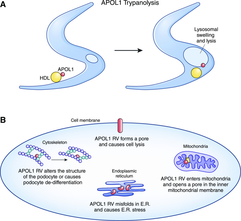
Rates of many types of severe kidney disease are much higher in Black individuals than most other ethnic groups. Much of this disparity can now be attributed to genetic variants in the apoL1 (APOL1) gene found only in individuals with recent African ancestry. These variants greatly increase rates of hypertension-associated ESKD, FSGS, HIV-associated nephropathy, and other forms of nondiabetic kidney disease. We discuss the population genetics of APOL1 risk variants and the clinical spectrum of APOL1 nephropathy. We then consider clinical issues that arise for the practicing nephrologist caring for the patient who may have APOL1 kidney disease.

Keywords: APOL1; APOL1 protein; African Americans; Apolipoprotein L1; Focal Segmental; Genetics; Glomerulosclerosis; HIV-Associated Nephropathy; Kidney Genomics Series; Population; chronic kidney disease; diabetes mellitus; genetic kidney disease; human; hypertension; kidney.


Figures

Figure 1.
Figure 1.
APOL1 protein and its variants. (Top panel) APOL1 domains are named according to their role in trypanolysis, the first known function of APOL1. APOL1 is the only member of the APOL family with a signal peptide (gray) in some splice isoforms that could enable cell export or trafficking to the cell membrane. The APOL1 risk variants are located near the C- terminus in the serum resistance antigen (SRA)–binding domain (light blue). The pore-forming domain shares features of bacterial colicin pore-forming toxins (purple). The function of the membrane-addressing domain is not clear but has been proposed as a pH-sensitive switch that facilitates membrane insertion (yellow). (Middle panel) Sequences of the G0, G1, and G2 APOL1 variants from amino acids 339–398. This domain forms a coiled-coil structure with a leucine zipper motif; the G1 and G2 variants likely alter the functional binding properties of this domain. G0 is tightly bound by the trypanosome virulence factor SRA. G1-SRA binding is weaker, whereas G2 exhibits almost no binding to SRA. (Bottom panel) The allele frequency of G1 is approximately 23%, and G2 is approximately 15%. Two risk alleles (one inherited from the mother and one inherited from the father) are required for the APOL1 high-risk genotype that greatly increases risk of kidney disease. The three different possible high-risk allele combinations are G1/G1, G2/G2, and G1/G2. In most instances, one risk allele has no effect or, at most, a small effect on kidney disease.
Figure 2.
Figure 2.
APOL1 mechanisms in health and disease. (A) APOL1 is the key trypanolytic factor of human serum, providing protection against African trypanosomes. APOL1 (red) circulating on HDL complexes (yellow) or in complexes with IgM is taken up by trypanosomes in the bloodstream. APOL1 transits to the trypanosomal lysosome and inserts in the membrane, causing lysosomal swelling and leading to the death of the trypanosome. (B) The normal function of APOL1 in human kidneys is not known. Risk variant (RV) APOL1 (red) appears to be cytotoxic to kidney cells when expressed at high levels. Investigators have provided evidence to support multiple different molecular mechanisms of disease. Although the podocyte is usually proposed as the predominantly affected cell type, different mechanisms in different cell types may explain clinical presentations of APOL1 kidney disease that vary from aggressive to slowly progressive, highly or minimally proteinuric, and inflammatory or noninflammatory. E.R., endoplasmic reticulum.
Figure 3.
Figure 3.
APOL1 risk variants increase the risk of many different types of kidney disease in Black individuals. APOL1 risk variants increase the risk of hypertension-attributed ESKD (H-ESKD; 7- to 11-fold), FSGS (17-fold), HIV-associated nephropathy (HIVAN; 29- to 89-fold), nondiabetic CKD (2- to 4-fold), and other kidney disease manifestations. APOL1 does not appear to affect risk for developing IgA nephropathy or diabetic nephropathy (but does have some influence on progression in patients with diabetes and proteinuric kidney disease). In general, studies tend to show large increases in risk when the trigger for APOL1 kidney disease is known (e.g., IFN-associated FSGS/glomerulopathy and HIV), with histologically defined lesions on kidney biopsy, with hard end points such as ESKD, and when the background rate of disease in a group is low. Effects tend to be smaller when disease is defined by a continuous variable such as eGFR or proteinuria, in the case of soft end points defined by an arbitrary threshold such as CKD, and when the background rate of disease is high (increasing age or among patients with diabetes). DKD, diabetic kidney disease; HR, high-risk; OR, odds ratio.
Figure 4.
Figure 4.
Donor APOL1 genotype determines graft survival in kidney transplantation. Transplants from APOL1 high-risk genotype donors (A) have decreased graft survival compared with kidneys from nonrisk donors. The recipient APOL1 genotype (B) does not appear to change the outcome for the survival of the donor kidney. This natural experiment indicates that it is probably kidney-expressed APOL1 rather than circulating APOL1 that injures in the kidney in the transplant setting, but it also suggests that kidney-expressed APOL1 may be the toxic factor when disease occurs in the native kidney. Some caution in making this extrapolation from transplant to native kidneys is warranted given the complex medical regimens of transplant recipients. It is not yet clear how the APOL1 genotype affects the outcome of kidney donors, but some studies suggest higher rates of GFR decline in APOL1 high-risk donors than in nonrisk donors. RV, risk variant.

References

    1. Genovese G, Friedman DJ, Ross MD, Lecordier L, Uzureau P, Freedman BI, Bowden DW, Langefeld CD, Oleksyk TK, Uscinski Knob AL, Bernhardy AJ, Hicks PJ, Nelson GW, Vanhollebeke B, Winkler CA, Kopp JB, Pays E, Pollak MR: Association of trypanolytic ApoL1 variants with kidney disease in African Americans. Science 329: 841–845, 2010. - PMC - PubMed
    1. Tzur S, Rosset S, Shemer R, Yudkovsky G, Selig S, Tarekegn A, Bekele E, Bradman N, Wasser WG, Behar DM, Skorecki K: Missense mutations in the APOL1 gene are highly associated with end stage kidney disease risk previously attributed to the MYH9 gene. Hum Genet 128: 345–350, 2010. - PMC - PubMed
    1. Friedman DJ, Pollak MR: Apolipoprotein L1 and kidney disease in African Americans. Trends Endocrinol Metab 27: 204–215, 2016. - PMC - PubMed
    1. Kruzel-Davila E, Wasser WG, Skorecki K: APOL1 nephropathy: A population genetics and evolutionary medicine detective story. Semin Nephrol 37: 490–507, 2017. - PubMed
    1. Smith EE, Malik HS: The apolipoprotein L family of programmed cell death and immunity genes rapidly evolved in primates at discrete sites of host-pathogen interactions. Genome Res 19: 850–858, 2009. - PMC - PubMed

Publication types