. 2020 Aug 14;369(6505):818-823.
doi: 10.1126/science.abc5343. Epub 2020 Jul 2.

Primary exposure to SARS-CoV-2 protects against reinfection in rhesus macaques

Affiliations

Primary exposure to SARS-CoV-2 protects against reinfection in rhesus macaques

Wei Deng et al. Science. .

Abstract

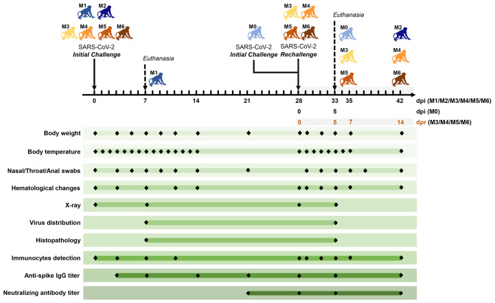
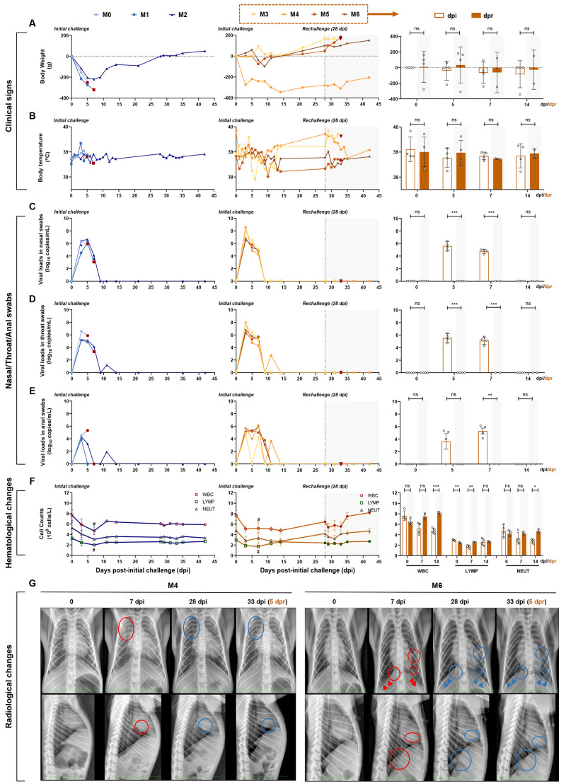
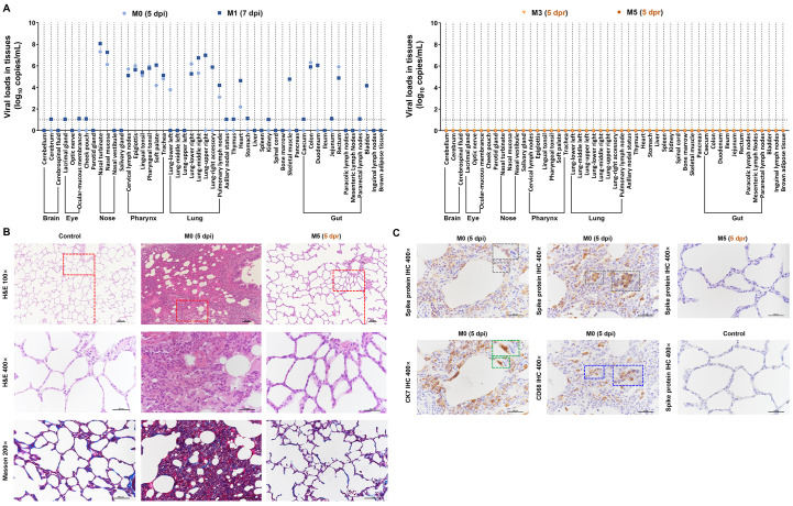
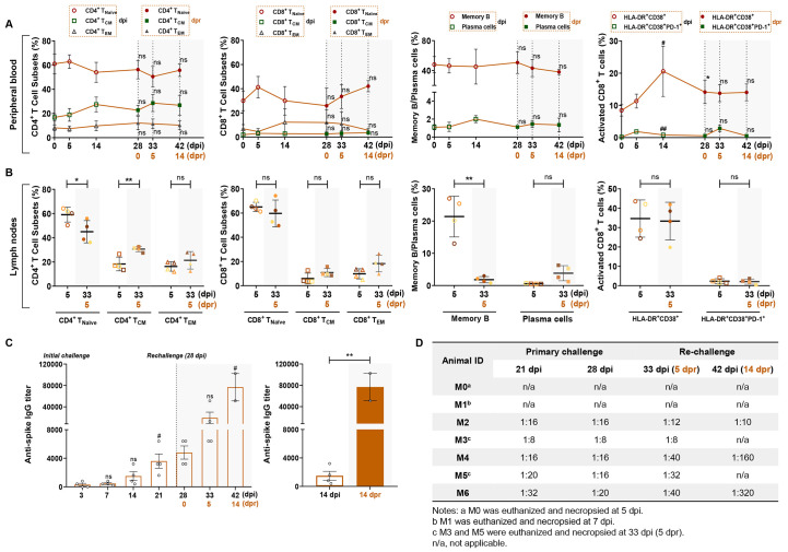
Coronavirus disease 2019 (COVID-19), which is caused by infection with the severe acute respiratory syndrome coronavirus 2 (SARS-CoV-2), has become a global pandemic. It is unclear whether convalescing patients have a risk of reinfection. We generated a rhesus macaque model of SARS-CoV-2 infection that was characterized by interstitial pneumonia and systemic viral dissemination mainly in the respiratory and gastrointestinal tracts. Rhesus macaques reinfected with the identical SARS-CoV-2 strain during the early recovery phase of the initial SARS-CoV-2 infection did not show detectable viral dissemination, clinical manifestations of viral disease, or histopathological changes. Comparing the humoral and cellular immunity between primary infection and rechallenge revealed notably enhanced neutralizing antibody and immune responses. Our results suggest that primary SARS-CoV-2 exposure protects against subsequent reinfection in rhesus macaques.


Figures

Fig. 1
Fig. 1. Experimental design and sample collection.
Seven adult Chinese-origin rhesus macaques (M0 to M6) were enrolled in the current study. At the outset of this experiment, six monkeys (M1 to M6) were challenged intratracheally with SARS-CoV-2 at 1 × 10⁶ TCID50. After all the experimentally-infected monkeys had recovered from the primary infection, four infected monkeys (M3 to M6) were intratracheally rechallenged at 28 days post initial challenge (dpi) with the same dose of the SARS-CoV-2 strain, to ascertain the possibility of reinfection. In addition, an uninfected monkey (M0) was also treated with SARS-CoV-2 as the model control of the second challenge, and a previously infected monkey (M2) was untreated in the rechallenge experiment and was continuously monitored as the control animal. To compare the virus distribution and histopathological changes between the initially infected monkeys and the reinfected monkeys, two monkeys per group (M0 and M1 in the initial infection group, M3 and M5 in the reinfection group) were euthanized and necropsied at 5 dpi (M0), 7 dpi (M1) and 5 days post rechallenge (dpr) (M3 and M5), respectively. Body weight, body temperature, nasal/throat/anal swabs, hematological changes, immunocytes, and specific antibodies were measured over the short-term observation period. Two measurements of virus distribution and histopathology (H&E/IHC staining) were carried out at 5 dpi (M0), 7 dpi (M1), and 5 dpr (M3 and M5). Chest X-ray and neutralizing antibody titers against SARS-CoV-2 were examined at the indicated time points.
Fig. 2
Fig. 2. Longitudinal tracking of clinical signs, viral replication, hematological changes, and radiological changes.
(A and B) Clinical signs in each monkey. Monkeys were examined daily for changes in body weight and rectal temperature over the observation period after the initial infection, followed by virus rechallenge. The changes in weight are expressed as body weight loss prior to primary infection. (C, D, and E) Detection of viral RNA in nasal, throat, and anal swabs. The SARS-CoV-2 RNA was detected by qRT–PCR in the swabs from seven monkeys at the indicated time points. (F) Hematological changes, including WBC, LYMP, and NEUT counts in the peripheral blood, were monitored. (G) Chest X-rays of animals at 0, 7, 28, and 33 dpi (5 dpr) were examined and the representative images of M4 and M6 are shown (red circles, areas of interstitial infiltration and exudative lesion; red arrows, obscured diaphragmatic surface; blue circles and arrows, areas that have recovered from pneumonia). Four monkeys (M3 to M6) were rechallenged at 28 dpi (dotted line and shaded areas), and the results of the initial infection and rechallenge were compared in bar graphs. The bars represent the average of four rechallenged animals at the indicated time points. The viral RNA in nasal, throat, and anal swabs of rechallenged animals were significantly lower than those of the initial infection, while significant hematological changes were observed between the primary and second challenges (unpaired t-test, dpi vs. dpr, *P < 0.05; **P < 0.01; ***P < 0.001; #P < 0.05 0 dpi vs. 7 dpi).
Fig. 3
Fig. 3. Comparison of virus distribution and pathological changes between the primary challenge and rechallenge stages.
(A) Detection of viral RNA in the indicated organs (brain, eye, nose, pharynx, lung, and gut. Compared with M0 and M1 (at 5 or 7 dpi; primary infection stage), viral replication tested negative in the indicated tissues from M3 and M5 (at 5 dpr; virus rechallenge stage). Using a viral load > 10 log10 copies/mL as the threshold of positivity-tissue-based PCR, tissues from 49 anatomical parts were detected for qualifying virus-infected positivity. Fourteen tissues from the respiratory tract, gut, and heart exhibited SARS-CoV-2-positive cells in both M0 and M1. SARS-CoV-2-positive cells were only observed in the left lower lung from M0 or in the right upper lung, upper accessory lung, skeletal muscle, duodenum, and bladder from M1. The remaining tissues from 29 anatomical parts did not show SARS-CoV-2-positive cells, indicating that these tissues were intact from viral invasion. (B) In M0 (5 dpi), an interstitial lesion including remarkedly widened alveolar septa and massive infiltrated inflammatory cells was observed using H&E staining. A mild fibrosis was clearly detected within widened alveolar septa using Masson staining. IHC against the spike protein of SARS-CoV-2 (7D2, grey frame), macrophages (CD68, blue frame), or alveolar epithelial cells (CK7, green frame) are visualized in parallel in Fig. 3C. The spike-positive cells overlapped with either alveolar epithelial cells or macrophages showing diffused interstitial pneumonia affected by SARS-CoV-2 invasion. In M5 (5 dpr), no remarked pathological changes and virus distribution were detected via H&E staining, Masson staining, or IHC, indicating that the interstitial lesions had completely recovered from the SARS-CoV-2 primary infection and were intact to reinfection. The red rectangles indicate the areas of magnification. Black scale bar at 100× or 200× = 100 μm. Black scale bar at 400× = 50 μm. Data are representative of three independent experiments.
Fig. 4
Fig. 4. Comparison of cellular and humoral immunity between primary challenge and rechallenge stages in macaques.
Four macaques (M3–M6) were rechallenged at 28 dpi (dotted line and shaded areas), and the results of the initial infection and rechallenge were compared at the same time points after the challenge and after the rechallenge. (A) Percentages of memory CD4+/CD8+ T cell subsets, memory B cells, plasma cells, or activated CD8+ T cells from peripheral blood for the challenge–rechallenge experiments. Compared with 0, 5 or 14 dpi, there was no significant differences on the percentage of naive CD4+/CD8+ T cells (CD4+/CD8+ Tnaïve, CD3+ CD4+/CD8+ CCR7+ CD45RA+), central memory CD4+/CD8+ T cells (CD4+/CD8+ Tcm, CD3+ CD4+/CD8+ CCR7+ CD45RA), effective memory T cells (CD4+/CD8+ Tem, CD3+ CD4+/CD8+ CCR7- CD45RA), memory B cells (CD3 CD20+ CD27+) and plasma cells (CD3 CD20+ CD43+) from peripheral blood at 0, 5, 14 dpr (unpaired t-test, ns, P > 0.05). The activation of CD8+ T cells (CD8+ CD38+ HLA-DR+ or CD8+ CD38+ HLA-DR+ PD-1+) at 14 dpi were increased compared with the baseline (unpaired t-test, #P < 0.05, ##P < 0.01), and elevated levels of CD8+ CD38+ HLA-DR+ T cells were also observed at 28 dpi (unpaired t-test, 0 dpi vs 0 dpr, * P < 0.05). (B) Percentages of memory CD4+/CD8+ T cell subsets, memory B cells, plasma cells or activated CD8+ T cells from lymph nodes between 5 dpi and 5 dpr. An increased percentage of CD4+ Tcm cells and decreased percentage of CD4+ Tnaïve cells and memory B cells from lymph nodes were found in the dot plots (unpaired t-test, *P < 0.05, **P < 0.01). (C) Levels of specific IgG against the spike protein of SARS-CoV-2 in four rechallenged monkeys. The levels of anti-viral antigen-specific IgG from each monkey were detected at 3, 7, 14, 21, 28, 33, and 42 dpi. Significantly increased levels of IgG were observed between the primary and second challenges (unpaired t-test, **P < 0.01, 14 dpi vs. 14 dpr; # P < 0.05, 3 dpi vs. 21 dpi, 28 dpi vs. 42 dpi). (D) Neutralizing antibody titers for protection of SARS-CoV-2-infected monkeys against reinfection.

Comment in

  • Can COVID-19 strike twice?
    York A. York A. Nat Rev Microbiol. 2020 Sep;18(9):477. doi: 10.1038/s41579-020-0424-x. Nat Rev Microbiol. 2020. PMID: 32690876 Free PMC article.

Similar articles

  • SARS-CoV-2 infection protects against rechallenge in rhesus macaques.
    Chandrashekar A, Liu J, Martinot AJ, McMahan K, Mercado NB, Peter L, Tostanoski LH, Yu J, Maliga Z, Nekorchuk M, Busman-Sahay K, Terry M, Wrijil LM, Ducat S, Martinez DR, Atyeo C, Fischinger S, Burke JS, Slein MD, Pessaint L, Van Ry A, Greenhouse J, Taylor T, Blade K, Cook A, Finneyfrock B, Brown R, Teow E, Velasco J, Zahn R, Wegmann F, Abbink P, Bondzie EA, Dagotto G, Gebre MS, He X, Jacob-Dolan C, Kordana N, Li Z, Lifton MA, Mahrokhian SH, Maxfield LF, Nityanandam R, Nkolola JP, Schmidt AG, Miller AD, Baric RS, Alter G, Sorger PK, Estes JD, Andersen H, Lewis MG, Barouch DH. Chandrashekar A, et al. Science. 2020 Aug 14;369(6505):812-817. doi: 10.1126/science.abc4776. Epub 2020 May 20. Science. 2020. PMID: 32434946 Free PMC article.
  • DNA vaccine protection against SARS-CoV-2 in rhesus macaques.
    Yu J, Tostanoski LH, Peter L, Mercado NB, McMahan K, Mahrokhian SH, Nkolola JP, Liu J, Li Z, Chandrashekar A, Martinez DR, Loos C, Atyeo C, Fischinger S, Burke JS, Slein MD, Chen Y, Zuiani A, Lelis FJN, Travers M, Habibi S, Pessaint L, Van Ry A, Blade K, Brown R, Cook A, Finneyfrock B, Dodson A, Teow E, Velasco J, Zahn R, Wegmann F, Bondzie EA, Dagotto G, Gebre MS, He X, Jacob-Dolan C, Kirilova M, Kordana N, Lin Z, Maxfield LF, Nampanya F, Nityanandam R, Ventura JD, Wan H, Cai Y, Chen B, Schmidt AG, Wesemann DR, Baric RS, Alter G, Andersen H, Lewis MG, Barouch DH. Yu J, et al. Science. 2020 Aug 14;369(6505):806-811. doi: 10.1126/science.abc6284. Epub 2020 May 20. Science. 2020. PMID: 32434945 Free PMC article.
  • Infection with novel coronavirus (SARS-CoV-2) causes pneumonia in Rhesus macaques.
    Shan C, Yao YF, Yang XL, Zhou YW, Gao G, Peng Y, Yang L, Hu X, Xiong J, Jiang RD, Zhang HJ, Gao XX, Peng C, Min J, Chen Y, Si HR, Wu J, Zhou P, Wang YY, Wei HP, Pang W, Hu ZF, Lv LB, Zheng YT, Shi ZL, Yuan ZM. Shan C, et al. Cell Res. 2020 Aug;30(8):670-677. doi: 10.1038/s41422-020-0364-z. Epub 2020 Jul 7. Cell Res. 2020. PMID: 32636454 Free PMC article.
  • Early Insights into Immune Responses during COVID-19.
    St John AL, Rathore APS. St John AL, et al. J Immunol. 2020 Aug 1;205(3):555-564. doi: 10.4049/jimmunol.2000526. Epub 2020 Jun 8. J Immunol. 2020. PMID: 32513850 Review.
  • The dynamics of humoral immune responses following SARS-CoV-2 infection and the potential for reinfection.
    Kellam P, Barclay W. Kellam P, et al. J Gen Virol. 2020 Aug;101(8):791-797. doi: 10.1099/jgv.0.001439. J Gen Virol. 2020. PMID: 32430094 Free PMC article. Review.

Cited by

References

    1. Zhu N., Zhang D., Wang W., Li X., Yang B., Song J., Zhao X., Huang B., Shi W., Lu R., Niu P., Zhan F., Ma X., Wang D., Xu W., Wu G., Gao G. F., Tan W.; China Novel Coronavirus Investigating and Research Team , A Novel Coronavirus from Patients with Pneumonia in China, 2019. N. Engl. J. Med. 382, 727–733 (2020). 10.1056/NEJMoa2001017 - DOI - PMC - PubMed
    1. “Coronavirus disease 2019 (COVID-19) Situation Report,” WHO. (2020). https://www.who.int/emergencies/diseases/novel-coronavirus-2019/situatio...
    1. Lan L., Xu D., Ye G., Xia C., Wang S., Li Y., Xu H., Positive RT-PCR Test Results in Patients Recovered From COVID-19. JAMA 323, 1502–1503 (2020). 10.1001/jama.2020.2783 - DOI - PMC - PubMed
    1. Zhou L., Liu K., Liu H. G., [Cause analysis and treatment strategies of “recurrence” with novel coronavirus pneumonia (COVID-19) patients after discharge from hospital]. Zhonghua Jie He He Hu Xi Za Zhi 43, 281–284 (2020). - PubMed
    1. J. An et al., Clinical characteristics of the recovered COVID-19 patients with re-detectable positive RNA test. medRxiv 2020.03.26.20044222 [Preprint]. 30 March 2020. .10.1101/2020.03.26.20044222 - DOI

Publication types

MeSH terms