. 2020 Jul 2;10(1):10833.
doi: 10.1038/s41598-020-67532-2.

Variability and sex-dependence of hypothermic neuroprotection in a rat model of neonatal hypoxic-ischaemic brain injury: a single laboratory meta-analysis

Affiliations

Variability and sex-dependence of hypothermic neuroprotection in a rat model of neonatal hypoxic-ischaemic brain injury: a single laboratory meta-analysis

Thomas R Wood et al. Sci Rep. .

Abstract

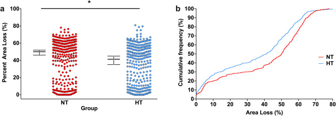
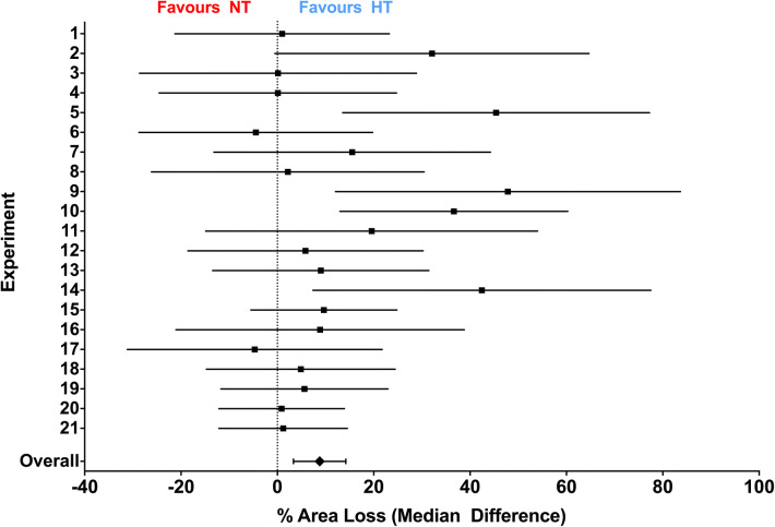
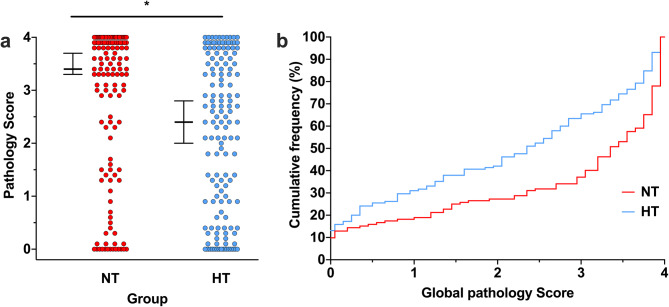
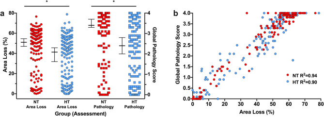
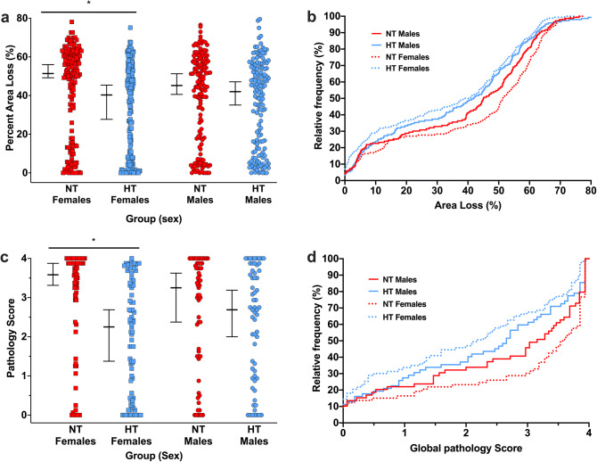
Therapeutic hypothermia (HT) is standard care for term infants with hypoxic-ischaemic (HI) encephalopathy. However, the efficacy of HT in preclinical models, such as the Vannucci model of unilateral HI in the newborn rat, is often greater than that reported from clinical trials. Here, we report a meta-analysis of data from every experiment in a single laboratory, including pilot data, examining the effect of HT in the Vannucci model. Across 21 experiments using 106 litters, median (95% CI) hemispheric area loss was 50.1% (46.0-51.9%; n = 305) in the normothermia group, and 41.3% (35.1-44.9%; n = 317) in the HT group, with a bimodal injury distribution. Median neuroprotection by HT was 17.6% (6.8-28.3%), including in severe injury, but was highly-variable across experiments. Neuroprotection was significant in females (p < 0.001), with a non-significant benefit in males (p = 0.07). Animals representing the median injury in each group within each litter (n = 277, 44.5%) were also analysed using formal neuropathology, which showed neuroprotection by HT throughout the brain, particularly in females. Our results suggest an inherent variability and sex-dependence of the neuroprotective response to HT, with the majority of studies in the Vannucci model vastly underpowered to detect true treatment effects due to the distribution of injury.


Conflict of interest statement

The authors declare no competing interests.

Figures

Figure 1
Figure 1
Hypothermic neuroprotection. (a) Scatter plot of percent area loss in the NT (n = 305) and HT (n = 317) groups. The median with 95% CI is plotted adjacent to each scatter. Across the 21 experiments, median (95% CI) area loss in was 50.1% (46.0–51.9%; n = 305) in the NT group, and 41.3% (35.1–44.9%; n = 317) in the HT group. *Denotes significant neuroprotection (p < 0.001). (b) Cumulative frequency plot from the NT (n = 305) and HT (n = 317) groups, with each observation resulting in a proportional increase in the y-axis. A relatively uniform degree of neuroprotection is seen across the whole range of injury severity, particularly between 10 and 60% area loss.
Figure 2
Figure 2
Median difference in area loss between NT and HT. Median difference (with 95% CI) in area loss between the NT and HT groups in each individual experiment, and overall. Experiments are numbered in chronological order. Of the 21 experiments, 15 favoured HT, and the overall median difference (absolute reduction in area loss in the HT group) was 8.8% (3.4–14.2%).
Figure 3
Figure 3
Global pathology scoring. (a) In the subset of animals that underwent formal pathology scoring (n = 277; n = 132 NT, n = 145 HT), median (95% CI) global pathology score 3.4 (3.3–3.7) in the NT group, and 2.4 (2.0–2.8) in the HT group. *Denotes significant neuroprotection (p < 0.001). (b) Cumulative frequency plot from the NT (n = 132) and HT (n = 145) groups, with each observation resulting in a proportional increase in the y-axis. A relatively uniform degree of neuroprotection is seen across the whole range of injury severity, with increasing relative neuroprotection in the HT group up to a global pathology score of 3.0.
Figure 4
Figure 4
Comparison between area loss and global pathology score. (a) Direct comparison of area loss and pathology score in the n = 277 (n = 132 NT, n = 145 HT) animals that underwent pathology scoring, representing the median injury in the NT and HT groups within each litter from each experiment. Median (95% CI) area loss was 51.0% (47.0–54.6%) in the NT group, and 41.3% (31.6–45.6%) in the HT group, with corresponding global pathology scores of 3.4 (3.3–3.7) in the NT group, and 2.4 (2.0–2.8) in the HT group. This resulted in a 19.0% neuroprotection by HT, compared to 25% neuroprotection when comparing the same animals using global pathology score. The resulting ES for HT was 0.42 when using area loss, and 0.51 when using global pathology. (b) Global pathology score was linearly correlated with area loss in both the NT (R2 = 0.94, p < 0.001) and HT (R2 = 0.90) groups (p < 0.001 for both).
Figure 5
Figure 5
Sex effects of hypothermic neuroprotection. (a) Median (95% CI) area loss in the NT group was 51.4% (49.2–56.0%; n = 159) in females, and males 45.2% (40.6–51.3%; n = 146) in males. In the HT group, median area loss was 40.3% (27.7–45.4%; n = 170) in females, and 42.0% (35.1–47.1%; n = 147) in males. The median differences between the NT and HT groups were 11.1% (21.6% neuroprotection) in females, and 3.2% (7.1% neuroprotection) in males. (b) Cumulative frequency distribution plot of area loss. (c) Median (95% CI) global pathology score in females was 3.6 (3.3–3.9, n = 73) in the NT group, and 2.3 (1.4–2.7, n = 83) in the HT group. In males, median global pathology score was 3.3 (2.4–3.6, n = 59) in the NT group, and 2.7 (2.0–3.2, n = 62) in the HT group. (d) Cumulative frequency global pathology score. The cumulative frequency plots suggest HT is neuroprotective across the entire range of injury in females, with neuroprotection in males centred solely around moderate injury scores (~ 20–55% area loss). *Denotes significant neuroprotection (p < 0.001).

References

    1. Franco NH. Animal experiments in biomedical research: a historical perspective. Animals (Basel). 2013;3(1):238–273. doi: 10.3390/ani3010238. - DOI - PMC - PubMed
    1. Pound P, Bracken MB. Is animal research sufficiently evidence based to be a cornerstone of biomedical research? BMJ. 2014;348:g3387. doi: 10.1136/bmj.g3387. - DOI - PubMed
    1. Jacobs SE, Berg M, Hunt R, Tarnow-Mordi WO, Inder TE, Davis PG. Cooling for newborns with hypoxic ischaemic encephalopathy. Cochrane Database Syst. Rev. 2013;1:CD003311. doi: 10.1002/14651858.CD003311.pub3. - DOI - PMC - PubMed
    1. Crossley NA, Sena E, Goehler J, Horn J, van der Worp B, Bath PM, Macleod M, Dirnagl U. Empirical evidence of bias in the design of experimental stroke studies: a metaepidemiologic approach. Stroke. 2008;39(3):929–934. doi: 10.1161/STROKEAHA.107.498725. - DOI - PubMed
    1. Shankaran S, Laptook AR, Pappas A, McDonald SA, Das A, Tyson JE, Poindexter BB, Schibler K, Bell EF, Heyne RJ, Pedroza C, Bara R, Van Meurs KP, Huitema CMP, Grisby C, Devaskar U, Ehrenkranz RA, Harmon HM, Chalak LF, DeMauro SB, Garg M, Hartley-McAndrew ME, Khan AM, Walsh MC, Ambalavanan N, Brumbaugh JE, Watterberg KL, Shepherd EG, Hamrick SEG, Barks J, Cotten CM, Kilbride HW, Higgins RD, Eunice Kennedy Shriver National Institute of Child H, Human Development Neonatal Research N Effect of depth and duration of cooling on death or disability at age 18 months among neonates with hypoxic–ischemic encephalopathy: a randomized clinical trial. Jama. 2017;318(1):57–67. doi: 10.1001/jama.2017.7218. - DOI - PMC - PubMed

Substances